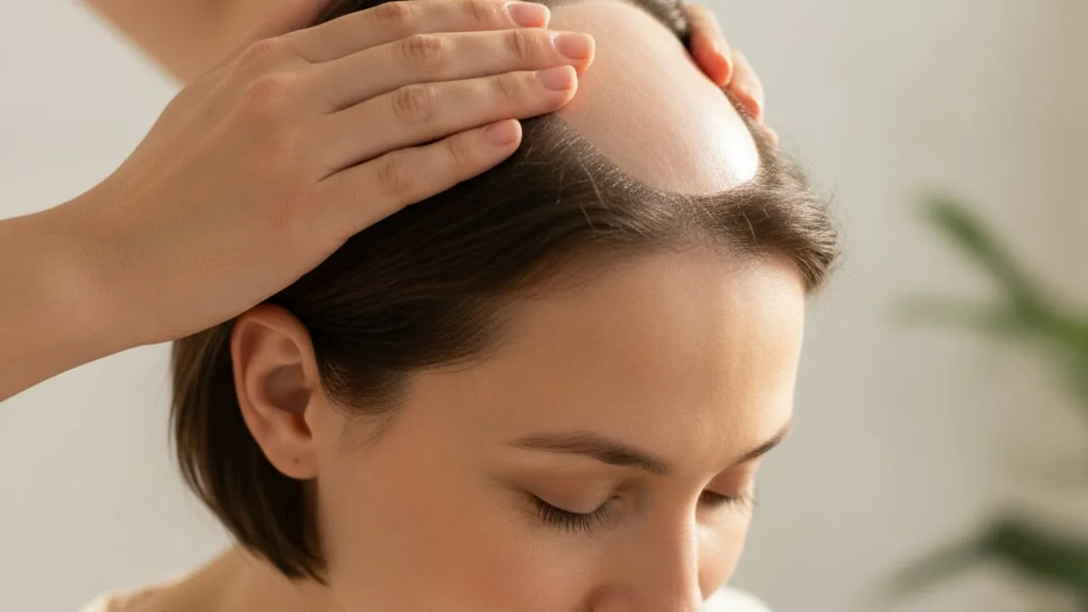
Bald Spot on the Back of Your Head: Causes and What Helps
What follows covers how crown thinning is graded, the other causes a clinician considers, the signs that deserve prompt attention, which labs are worth raising, and the kinds of options a licensed provider may discuss after an evaluation.
What does a thin or bare area at the crown usually mean?
It usually means androgenetic alopecia showing up at the vertex, the whorl at the back and top of the head. Research suggests that androgenetic alopecia is the most common form of hair loss and involves progressive miniaturization of hair follicles in a patterned distribution 1.
Miniaturization is the key idea. Follicles are not dying off all at once and leaving a clean hole. With each growth cycle, susceptible follicles produce a hair that is slightly shorter, finer and less pigmented than the one before. The density of follicles changes slowly, but the mass of hair they produce falls quickly enough that light starts passing through to the scalp. That is why crown loss is so often noticed in a photograph taken from behind, in a barber's second mirror, or in harsh overhead light, long before it is obvious in a bathroom mirror.
The shape matters too. Pattern loss at the crown tends to be a diffuse, soft-edged zone of thinner hair that spreads outward from the whorl, with hairs of visibly different thicknesses inside it. A true bald spot, meaning a sharply bordered area where the skin is smooth and bare, is a different clinical picture and points elsewhere.
None of this is cosmetic trivia to the person it is happening to. Wanting a clear answer early is reasonable, not vanity.
Why does the crown thin before the sides and back?
Because follicles in different scalp regions respond differently to androgens. Inside susceptible follicles, testosterone is converted by the enzyme 5-alpha-reductase into Dihydrotestosterone, a more potent androgen, and research suggests that this local androgen signalling drives the follicle miniaturization seen in androgenetic alopecia 3.
The follicles on the crown and the mid scalp carry the genetic susceptibility that makes them respond this way. The follicles along the lower back and sides of the head generally do not, which is why most men keep a rim of hair even after decades of crown loss, and why surgical restoration takes follicles from that rim.
Androgens are not the whole story. Studies suggest that other local factors around the follicle, including inflammation and oxidative stress, contribute to the process alongside androgen signalling 4. That helps explain why two people with similar hormone results can have very different scalps.
It also explains a common source of confusion. A blood hormone level and what is happening inside a follicle are not the same thing, so a single hormone result cannot confirm or exclude patterned thinning on its own. The diagnosis is made by looking at the scalp, not by reading one number.
Is crown thinning the same thing as male pattern hair loss?
Crown thinning is one presentation of male pattern hair loss, not a separate condition. In men, the patterned form commonly combines recession at the temples with thinning at the vertex, in varying proportions and in varying order 5.
Some men recede at the front first and keep a dense crown for years. Others hold a straight hairline while the vertex opens up, which is the version that arrives as a surprise, because the front view in the mirror looks unchanged. A third group thins in both areas at once until the two zones meet. All three are the same underlying process expressed differently.
If you want the wider picture of how the pattern develops, who it affects and what the course tends to look like, the guide to male pattern hair loss covers it in more detail than a crown-focused page can.
Can women develop a thin area at the crown too?
Yes. Androgenetic alopecia affects women as well as men, and the female presentation is usually diffuse thinning across the top and crown rather than a receding hairline 1.
The classic description is a widening of the central part, sometimes compared to a Christmas tree shape, with the frontal hairline largely preserved. Hair feels thinner in the hand, the ponytail circumference drops, and the scalp becomes visible along the part under bright light. The crown can be involved heavily enough that a thin patch is obvious when the hair is lifted or when it is wet.
A sharply defined bare circle in a woman is less typical of patterned loss, and a clinician would consider the other causes below before settling on that label. Shedding after a major illness, after a crash in energy intake, or alongside thyroid or iron issues can also show up as a thinner crown, and the two processes can overlap in the same person.
How do clinicians describe and grade thinning at the vertex?
With a pattern classification, standardized photographs and a close look at the scalp surface. For men, the Hamilton-Norwood classification describes the stage and distribution of patterned loss, including vertex involvement 5. For women, the Ludwig and Sinclair scales describe the degree of central and crown thinning and the width of the part.
The grade itself is less useful than what it is for. A stage recorded today, with photographs taken from the same angles and in the same light, gives a baseline that any future comparison is measured against. Hair changes slowly and memory is unreliable, so an honest before-and-after is worth more than an impression.
Alongside the grade, a clinician typically records part width, whether miniaturized hairs are visible mixed in with terminal hairs, how much scalp shows under direct light, and whether the back and sides are involved. Involvement of the lower occipital scalp is unusual in patterned loss and is one of the details that pushes the assessment in another direction.
What else can cause a bare patch at the back of the scalp?
Several conditions can produce a patch at the back of the head, and the differential includes patterned loss, telogen effluvium, alopecia areata, scalp infection, traction and the scarring alopecias 6. The table below sets out how they tend to present. Appearances overlap, and only an in-person examination can sort them out.
| Cause | How the area tends to look | Scalp surface | Typical course |
|---|---|---|---|
| Pattern hair loss at the crown | Diffuse, soft-edged thinning around the whorl, hairs of mixed thickness | Normal skin, follicular openings still visible | Gradual over years |
| Alopecia areata | Sharply bordered round or oval patch, sometimes several | Smooth, normal-coloured, openings preserved | Appears over weeks, course varies |
| Telogen effluvium | Thinning all over, often most obvious at the crown and part | Normal, with increased shedding when hair is washed or combed | Follows an illness, surgery, major stress or nutritional change, by months |
| Traction alopecia | Thin band or zone where hair has been pulled tightly | Normal early, may show small bumps or broken hairs | Builds with repeated styling tension |
| Scarring (cicatricial) alopecias | Patch that may be shiny or irregular, hairs sometimes emerging in tufts | Redness, scale, pustules, pain or itch, loss of visible follicular openings | Can progress steadily, follicles lost permanently |
| Scalp fungal infection | Scaly patch with broken hairs, more common in children | Scale, crust, sometimes a boggy swollen area | Spreads without care, contagious |
| Pressure-related loss | Patch at the point of prolonged contact, often at the back | Normal or tender | Follows long immobility or prolonged pressure |
| Hair pulling | Irregular outline, hairs broken at different lengths | Normal skin, no scale | Fluctuates, often linked to stress |
How does a clinician work out which one it is?
By combining the story with a close examination of the scalp rather than by guessing from a photograph. Evaluation for hair loss starts with history and physical examination, with targeted testing added when the history or examination suggests a contributing cause 6.
The history covers how fast it appeared, whether hair is shedding or simply thinner, family history on both sides, recent illness, surgery, weight change or restrictive eating, iron status, thyroid symptoms, scalp symptoms such as itch or pain, hairstyling habits, and a review of recent medicines and supplements.
The examination covers the whole scalp, not only the patch. A clinician looks at the hairline, the part, the crown and the occipital scalp, checks whether follicular openings are still visible, looks for redness, scale or pustules, and performs a gentle hair pull test at several sites to gauge active shedding. Eyebrows, lashes, body hair and nails are often checked too, because some conditions touch more than the scalp.
From there, the usual additions are trichoscopy, sometimes a scalp swab or scraping when infection is possible, and a small scalp biopsy when the picture is unclear or scarring is suspected. Blood tests are selective rather than routine, and they support the diagnosis rather than make it.
What does trichoscopy show on the scalp?
Trichoscopy is dermoscopy of the scalp, a magnified look at hair shafts, follicular openings and the skin between them. Research suggests that trichoscopy may help distinguish scarring from nonscarring alopecia in the clinic and can guide where a biopsy is taken 7.
In patterned loss, the signature finding is hair diameter diversity, meaning thick, intermediate and very fine hairs sitting side by side in the same area, usually with more single-hair follicular units at the crown than at the back of the head. In alopecia areata, short broken hairs, tapered hairs and yellow dots are described within an otherwise smooth patch. In the scarring alopecias, the critical observation is the absence of follicular openings, sometimes with scale around the hair shafts or hairs emerging in tufts 7.
For the reader, the practical point is simple. A magnified scalp exam takes minutes, is painless, and answers questions that no amount of photographing your own crown will settle. It is also how a clinician decides whether a biopsy is needed at all.
What if your situation looks like one of these?
Different stories point in different directions. The patterns below are common reasons people search for an answer about the back of the head. None of them is a diagnosis, and each ends in the same place, which is an examination.
A smooth, round patch that appeared within weeks
A well-defined circle of bare, normal-looking skin that turned up quickly is the classic description of alopecia areata rather than patterned loss, especially when the skin is smooth and the follicular openings are still visible 6. It can appear anywhere, including the occipital scalp and the beard. The course is unpredictable, so an early clinician visit is worth arranging rather than waiting to see what the patch does.
Thinning that crept in over years with no clear patch
Gradual loss of density around the whorl, with the hair there looking finer and shorter than the hair at the sides, fits the patterned process described above. It is worth photographing now, from directly above and from behind, in consistent light. Those photos become the comparison point for any decision made later, and they are more informative than counting hairs in the sink.
Heavy shedding everywhere, a few months after an illness or a hard stretch
When hair comes out in handfuls across the whole scalp rather than from one spot, and the trigger sits a few months back, telogen effluvium is part of the differential a clinician considers 6. The crown often looks worst because that is where density was lowest to begin with. This type of shedding is also the one most often linked by a clinician to iron, thyroid or intake issues, which is where targeted blood tests earn their place.
A patch that is red, scaly, itchy or sore
Symptoms on the skin itself change the priority. Redness, scale, pustules, burning, tenderness or a patch where the pinpoint follicular openings have disappeared raise the possibility of an inflammatory or scarring process, and in scarring alopecias follicles can be lost permanently. This is the situation that is worth discussing with a clinician promptly rather than monitoring at home.
A thin band where hair is pulled tight
Thinning that follows the line of a tight ponytail, bun, braid, weave or helmet strap suggests tension on the follicles rather than hormonal miniaturization. Early traction loss can improve when the tension is removed, while long-standing traction can leave permanent gaps. A clinician can tell you which stage the area is in, which is the thing you cannot judge by eye.
Which signs mean talking to a clinician promptly?
Any scalp change that involves the skin, not just the hair, deserves prompt attention. Book an appointment sooner rather than later when you notice redness, scale, crusting or pustules in the area, pain, burning or persistent itch, a patch that is spreading quickly, hairs that snap off at the surface, a smooth patch where you can no longer see the tiny follicular openings, or hair loss arriving together with other changes such as marked fatigue, weight change, nail pitting or loss of eyebrow and body hair.
Sudden loss in a child, a swollen or boggy area, or a patch after a scalp injury also belong in the same category. The general principle holds for anything else you notice on your scalp. An abnormal finding is never worth ignoring, and a short visit is a reasonable price for knowing what you are looking at.
Why does an early answer matter more than it seems?
Because the window for action is different for each cause, and two of the categories are time-sensitive. In scarring alopecias, the follicular openings are destroyed, which is why trichoscopy specifically looks for their absence 7. Follicles that are already gone are not coming back, so the realistic aim in those conditions is to limit further loss, and that aim depends on recognising the process early.
Patterned loss works on a slower clock, but it is progressive, with follicles miniaturizing cycle after cycle 1. Decisions made while miniaturized follicles are still producing hair are made with more material to work with than decisions made after an area has gone bare.
There is also a practical reason. An early visit gives you a documented baseline, a named cause, and a plan for what to watch. Without one, people spend years cycling through shampoos and supplements chosen for a condition they were never shown they have.
Which lab tests are worth raising with a provider?
Labs play a supporting role in hair loss, and they are chosen to match the history rather than ordered by reflex. Evaluation guidance for hair loss includes targeted laboratory testing when the history or examination suggests a contributing cause 6. The tests that most often come up in a scalp consultation are:
- Thyroid tests, since thyroid dysfunction can affect the hair cycle.
- Ferritin, as a marker of iron stores, which clinicians commonly review in people with diffuse shedding.
- DHT and related androgen markers, read in context with the scalp examination rather than on their own.
Depending on the history, a provider may add markers of overall nutritional and metabolic status, since restrictive dieting, very low energy intake and rapid weight loss all show up in the hair cycle months later. FitScript's membership includes 150+ biomarkers tested per year, with Atlas, the AI health advisor, writing the interpretation that a licensed provider then reviews.
One caution worth repeating. A normal panel does not rule out patterned loss, and an out-of-range result does not by itself explain a bald area. Numbers narrow the possibilities and point to contributing factors, and the scalp examination still carries the diagnosis.
What options may a licensed provider consider?
Options fall into a handful of broad classes, and which ones fit depends entirely on the diagnosis rather than on the size of the area. A licensed provider evaluates whether any of them is appropriate after examining your scalp and reviewing your history, and no article can make that call.
- Topical therapies applied directly to the scalp.
- Oral prescription therapies that act on the hormonal pathway behind patterned loss.
- In-office procedures, including injectable preparations made from your own blood and light-based or energy-based devices.
- Surgical restoration, which relocates follicles from the less androgen-responsive back and sides of the scalp.
- Anti-inflammatory approaches, which a provider may consider when the examination points to an inflammatory or scarring process.
- Cosmetic camouflage, such as fibres, pigments and scalp micropigmentation, which changes appearance without changing follicles.
Doses, schedules, combinations and duration are decisions for the provider who examined you, and any decision to start, stop or change a therapy belongs to that provider. A provider-led route is outlined on the hair loss treatment for men page. FitScript works with licensed providers across all 50 states; specific treatment availability can vary by state and is confirmed during your provider evaluation.
Set expectations on time as well. Hair grows slowly and the follicle cycle is measured in months, so any assessment of whether something is changing is made with standardized photographs over months, not with a week of close inspection in the mirror. No approach promises a particular result, and honest follow-up is the only way to judge.
What helps day to day while you are getting answers?
Sensible scalp habits will not undo miniaturization, but they stop you adding avoidable damage on top of it. The list is short and unglamorous.
Release the tension. Tight ponytails, buns, braids, weaves, extensions and tight headwear pull on follicles, and that mechanical load is the one cause of patchy loss you can reduce yourself today. Handle wet hair gently, since hair is at its most fragile then, and go easy on high heat, hard brushing and aggressive chemical processing while an area is already thin.
Feed the cycle. Hair follicles are metabolically demanding, and prolonged low energy intake, crash dieting and low protein intake tend to show up in the hair months later. Eat enough, eat enough protein, and address iron or thyroid issues with your clinician rather than guessing with supplements.
Protect exposed scalp from sun, since skin that used to be covered by hair now is not. Sleep properly and manage the stress load you can control, both of which are ordinary health advice rather than a hair remedy.
And take the photographs. Same spot, same light, once a month, from above and from behind. That habit costs nothing and will tell you and your provider more than anything else on this list.
Which common beliefs about crown balding are worth dropping?
Several. Hats do not cause patterned thinning, and neither does washing your hair regularly, though both get blamed because shed hairs become visible when you wash and because hats are worn by people who are already thinning.
Shaving does not change the thickness of the hair that grows back, since the shaft tapers naturally and a cut end simply feels blunter. Scalp massage and brushing will not restore follicles that are miniaturizing, however pleasant they feel. A thin crown does not mean your testosterone is unusually high, because the process depends on local follicle sensitivity rather than on an impressive blood level, which is why hormone results are read alongside the examination rather than instead of it.
Finally, a few extra hairs in the shower after a stressful month are not the same as a bald spot, and a bald spot that arrived in three weeks is not the same as patterned loss. The difference is visible on examination, and it changes what happens next.
Key takeaways
- A thin or bare area at the crown is most often androgenetic alopecia, which miniaturizes follicles gradually rather than removing them overnight.
- Crown follicles are sensitive to local androgen signalling in a way that the follicles on the back and sides generally are not.
- A sharply bordered, smooth patch, or one with redness, scale, pain or missing follicular openings, points away from patterned loss and is worth discussing with a clinician promptly.
- Diagnosis rests on the history and a scalp examination, usually with trichoscopy, and sometimes with a biopsy or a swab.
- Labs play a supporting role, with thyroid tests, ferritin and DHT the ones most often raised, and none of them replaces the scalp exam.
- Options span topical, oral, procedural, surgical and cosmetic classes, and a licensed provider decides what is appropriate after an evaluation.
This page is educational and is not medical advice. Discuss your results, your scalp findings and any treatment decision with a clinician or licensed provider who has examined you.
References
- Androgenetic alopecia. Nature Reviews. Disease Primers, 2025.
- Androgenetic Alopecia: A Review. The Nigerian Postgraduate Medical Journal, 2024.
- Research progress on the pathogenesis of androgenetic alopecia. European Journal of Dermatology, 2025.
- Male androgenetic alopecia. Anais Brasileiros de Dermatologia, 2025.
- Hair Loss: Diagnosis and Treatment. American Family Physician, 2024.
- The dermatoscope in the hair clinic: Trichoscopy of scarring and nonscarring alopecia. Journal of the American Academy of Dermatology, 2023.
Common questions
Is a bald spot at the crown always permanent?
Not always, and it depends entirely on the cause. Patterned thinning tends to progress slowly, while shedding after an illness or a tight hairstyle can improve once the trigger is removed. Loss from a scarring process behaves differently, because the follicular openings are lost. Only a scalp examination can tell you which situation you are in.
How can I tell a bald spot from the natural whorl at the back of my head?
Everyone has a whorl where the hair changes direction, and it naturally lets more light through to the scalp. The question is whether the hair in that area has become finer and shorter than the hair at the sides, and whether the area has widened over time. Monthly photographs from directly above and behind, in the same light, answer it better than a mirror does.
Do hats, frequent washing or shaving make crown balding worse?
No. Wearing a hat does not cause patterned thinning, washing simply makes already-shed hairs visible, and shaving does not change how thick the regrowing hair is. Tight styles that pull on the roots are a real cause of mechanical loss, so that is the habit worth changing.
Does thinning at the crown mean my testosterone is high?
Not necessarily. Patterned thinning depends on how sensitive your scalp follicles are to androgens locally, not on having an unusually high blood level. That is why a hormone result is read alongside a scalp examination rather than on its own, and why a normal result does not rule anything out.
Which blood tests are usually worth asking about for hair loss?
Thyroid tests, ferritin as a marker of iron stores, and DHT are the ones that come up most often in a scalp consultation. They are chosen to match your history rather than ordered automatically. These tests support the assessment, they do not replace the scalp examination that identifies the cause.
Can women get a bald area at the back of the head?
Yes. In women, patterned loss usually shows as a widening central part and diffuse thinning across the top and crown, with the hairline largely preserved. A sharply defined bare circle is less typical of the patterned form and is a good reason to see a clinician for an examination.
When should I stop watching and book an appointment?
Book promptly if the area is red, scaly, crusted, painful or itchy, if it is spreading quickly, if hairs are snapping off at the surface, or if the skin looks smooth with no visible follicular openings. The same applies if hair loss comes with fatigue, weight change or nail changes. Scalp changes involving the skin itself are the time-sensitive ones.
How long before I can tell whether anything is changing?
Hair grows slowly, so change is judged over months rather than weeks. The practical approach is standardized photographs taken monthly from the same angles in the same light, reviewed with your provider at follow-up. Any decision to start, stop or change a therapy belongs to the licensed provider who evaluated you.