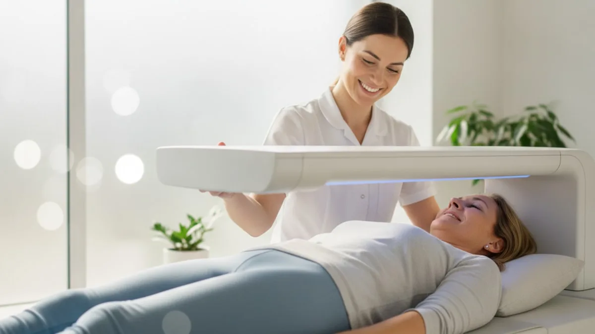
Bone Density Test (DEXA Scan): T-Scores Explained
The scan is short, involves no injection, and you stay dressed as long as your clothing has no metal. What the numbers mean depends on your age, your sex, which sites were measured, and whether anything inside the scanned area distorted the reading.
What does a bone density scan actually measure?
A bone density scan measures how much bone mineral sits inside a defined area of your skeleton, and reports it as areal bone mineral density in grams per square centimetre.
The word areal matters. The scanner sees a flat projection of a three dimensional bone, so it divides the mineral it detects by the area it passed over rather than by true volume 1. Two people with similar bone quality but different bone sizes can therefore produce different numbers, because a bigger bone presents more mineral within the same projected area. That is one reason a raw density value is rarely read on its own.
What appears on the report is the density value plus two comparison scores. The T-score compares your density with a young adult reference population, and the Z-score compares it with people of your own age and sex 2. Both are expressed in standard deviations, which is why they look like small positive or negative numbers rather than units of mass.
Density is a stand-in for bone strength, not a direct measurement of it. A bone resists breaking through its mineral content, the architecture of its internal lattice, its turnover rate and its geometry, and a scan captures the mineral part most directly. So the number is read next to your fracture history, your height, your age and the rest of your picture, not as a verdict by itself.
The test also answers a different question from blood work. Blood tests describe what is circulating right now. A scan describes what has accumulated in the skeleton over decades.
How does dual energy X-ray absorptiometry work?
Dual energy X-ray absorptiometry, written DXA and often spoken as DEXA, works by passing two different X-ray energies through the body and comparing how much of each is absorbed.
Bone and soft tissue absorb those two energies in different proportions, so the scanner software can separate the bone signal from everything around it and calculate the mineral content of what is left 1. A scanning arm travels above you while you lie on a padded table. Nothing touches you, nothing enters the body, and the room stays open.
Radiation exposure is low, and practice guidance still regards dose as something to keep as low as reasonably achievable, which is why scans are ordered to answer a defined question rather than out of curiosity 1.
Accuracy depends on the machine being kept honest. Services run regular quality control scans on a test object so that drift in calibration is spotted before it shows up in patient results, and that quality control history is what makes a scan from this year comparable with one from a previous year 1. A scanner that has not been checked against its own baseline cannot support reliable follow up.
Which parts of the skeleton get scanned, and why those?
Standard scans measure the lumbar spine and the upper part of the thigh bone, because those sites measure well and because fractures there carry real consequence.
The lumbar spine region usually covers the first through fourth lumbar vertebrae, and the hip is reported at the femoral neck and as the total hip 2. The forearm is kept as a fallback site, used when the spine or hip cannot be measured or analysed, or when body size exceeds what the scanner table can accommodate 2.
When several sites are measured, the classification follows the lowest valid score rather than an average of the sites, so a reassuring hip does not cancel out a low spine 2. Individual vertebrae are excluded from the spine calculation when they are structurally abnormal, for example after a compression fracture or where there is obvious local change, and a spine measurement needs at least two usable vertebrae to be reported at all 2.
The report should state which sites were measured, which levels were used or excluded, and why 1. If your report does not say, that is a fair question to raise, because the same skeleton can be classified differently depending on which levels went into the calculation.
What does a T-score mean?
A T-score says how far your bone mineral density sits from the average of a young adult reference population, counted in standard deviations.
A score of zero means you match that young adult average. A score of minus one means you sit one standard deviation below it. Because the scale is statistical rather than physical, small differences in the reference database used by the scanner can shift the score without anything changing in your bones.
| T-score, in words | Category used in reporting | What it describes |
|---|---|---|
| Minus one point zero or higher | Normal | Density sits within one standard deviation of the young adult average |
| Between minus one point zero and minus two point five | Low bone mass | Below the young adult average, above the osteoporosis threshold |
| Minus two point five or lower | Osteoporosis | At or below two and a half standard deviations under the young adult average |
These bands come from a standard classification, and they are applied to postmenopausal women and to men aged fifty and over 2. They are not applied in the same way to younger adults 2.
A band is a label on a continuum, not a line between health and illness. Fracture risk rises gradually as density falls, and a previous fracture from a minor fall carries weight in interpretation regardless of which band the score lands in. Only a clinician who knows your history can say what your band means for you.
What is a Z-score, and who is it for?
A Z-score compares your bone mineral density with people of the same age and sex rather than with young adults, which makes it the score that carries meaning in younger people.
For premenopausal women and for men under fifty, reports lean on the Z-score, because comparing a thirty year old skeleton with a young adult reference says little that is useful 2. A Z-score of minus two point zero or lower is described in reporting conventions as below the expected range for age, which is a flag for further thought rather than a label 2.
That flag usually prompts a clinician to look for contributors outside the skeleton, since low density for age in a younger adult often traces to something that can be identified and addressed. Hormonal status, digestive absorption, long periods of low energy availability in athletes, inflammatory conditions and some medicines can all sit behind a low Z-score. Ask your prescriber about anything you take, since only the prescriber can judge its relevance.
Older adults get both scores on the report. The T-score drives the category, and the Z-score adds context about whether the result is what age alone would predict or something more unusual 2.
Who should think about getting scanned?
Screening is aimed at people whose result would change a decision, and the clearest case is postmenopausal women at older ages.
Screening guidance summarised in an expert panel review supports measuring bone mineral density in women aged sixty five and older, and in younger postmenopausal women whose estimated risk, worked out with a formal risk assessment tool, is comparable to that of a sixty five year old woman 3. For men, the same review describes screening as less settled, because the data behind a population level recommendation have not been judged sufficient 3.
Beyond population screening, a scan is considered in adults who have broken a bone from a fall at standing height or less after the age of fifty, who have lost measurable height, who live with conditions that affect bone or mineral handling, or who take some medicines known to affect the skeleton over time 3. Again, that last group is a conversation with your prescriber, not something to work out alone.
Age, family history of hip fracture, low body weight, smoking, heavy alcohol intake and long periods of inactivity all feed into the decision. A clinician decides whether a scan is appropriate and when, and that decision is better than any self assessment from a symptom list, since low bone density produces no symptoms until something breaks.
What happens on the day of the scan?
You lie still on a padded table, fully clothed if your clothing has no metal, while a scanning arm passes above you for a few minutes.
There is no needle, no enclosed tunnel and no sedation. Metal inside the scanned area creates artefact, so zips, belt buckles, underwires, coins and jewellery around the hips and lower back are removed or swapped for a gown 5. Tests that use scan preparations can leave material in the abdomen that sits over the spine on the image, so those appointments are spaced apart from a bone scan and it is worth telling the clinic if you have had one recently 5.
Positioning is part of the measurement rather than a comfort step. For the spine, your lower legs are usually raised on a block so the lumbar curve flattens against the table. For the hip, your foot is held in a brace that rotates the leg inward so the femoral neck is presented at a consistent angle 5. Those positions are repeated at every follow up scan for the same reason.
Tell the technologist about previous fractures, spinal surgery, implants, scoliosis or a hip replacement before the scan starts, because those details change which regions can be analysed 5. The images are then read and reported, and your clinician goes through the result with you.
What can distort a result?
Anything inside the measured area that is not ordinary bone can push the number up or down, and most distortions push the spine number up.
At the spine, degenerative change in the joints and discs, calcification in the vessel running in front of the vertebrae, a collapsed vertebra, surgical hardware and marked curvature all add mineral to the projected area and inflate the apparent density 5. This is why an older spine can read higher than the hip in the same person, and why a reader excludes affected levels rather than reporting the spine as it falls 2.
At the hip, the errors are mostly about geometry. Rotating the leg too far in or out, letting the leg drift away from the midline, or placing the femoral neck box in the wrong spot all shift the value, and repeating those errors inconsistently between visits creates change that was never in the bone 6.
Analysis adds its own pitfalls. Mislabelling vertebral levels, selecting the wrong reference database, editing the bone outline by hand and accepting the software's automatic edges without checking them are recognised operator related errors, and training plus a consistent protocol is what keeps them out 6. If a result looks out of keeping with everything else about you, asking whether the images were reviewed for artefact is a reasonable question.
Why do two machines give two different numbers?
Scanners from different manufacturers are calibrated differently and carry different reference data, so an absolute density value from one machine does not transfer cleanly to another.
That difference is not a fault. It means a follow up scan belongs on the same machine wherever possible, and that switching services mid way through monitoring breaks the comparison unless a formal cross-calibration has been done between the two scanners 7. Guidance on monitoring treats cross-calibration as a prerequisite for comparing values produced by different equipment 7.
Even a single machine has a margin of its own. Repeat the same scan on the same person on the same day and the value will not be identical, and the size of that scatter is the precision error 7. Services calculate their own precision and convert it into a least significant change, the smallest difference between two scans that can be called real rather than noise.
The practical consequence is simple. A small drop between two scans is not automatically bone loss, and a small rise is not automatically improvement. Only a difference larger than the service's least significant change counts as a genuine change 7.
How often is it worth repeating?
A repeat scan is worth doing when enough time has passed that a real change could exceed the measurement margin, and when the result would change a decision.
Because bone mineral density moves slowly, the interval between scans is generally counted in years rather than months, and monitoring guidance frames the timing around whether expected change can clear the least significant change rather than around a fixed calendar rule 7. Scanning again too soon mostly measures the machine.
Several things shorten the interval in practice. A result near a decision threshold, a new fracture, a significant change in health, or starting something a clinician expects to affect bone can all justify an earlier look. Treatment decisions belong to a licensed provider, and the same provider sets the follow up interval that goes with them.
For comparison to be meaningful, the follow up scan should use the same machine, the same positioning protocol and the same regions of interest as the baseline, and the report should compare like with like rather than comparing a spine average that included four vertebrae with one that included three 17. Keep a copy of your baseline report, including the actual density values and not only the scores, so a future service can line them up correctly.
What does a low result say about fracture risk?
A low result raises estimated fracture risk, and that relationship is the whole reason the scan exists, but it describes probability rather than destiny.
Research suggests that bone mineral density measured by quantitative computed tomography is associated with vertebral fracture risk, and the same review reports a relationship for density measured by DXA 8. Lower density is associated with a higher likelihood of vertebral fracture across the populations examined 8.
Density is only one input. Risk calculators used alongside a scan combine the femoral neck density value with clinical factors such as age, sex, previous fracture, family history of hip fracture, smoking and alcohol intake to produce an estimated probability over a set period 3. Two people with identical T-scores can sit at quite different estimated risk once those factors are added.
The reverse also holds. A score inside the normal band does not rule out a future fracture, particularly in someone who falls often or who has already broken a bone from a minor fall, which is why fracture history is weighted heavily in interpretation 3. What your own number means for your own risk is a conversation with your clinician, not an arithmetic step.
What else can a DXA report show beyond bone mineral density?
Modern scanners produce several outputs from the same session, and density is only the headline one.
Research suggests that measures derived from the lumbar spine image, such as trabecular bone score, add information about bone texture beyond the density value alone 9. Studies indicate that a dedicated spine image acquired at the same visit can be used to look for vertebral fractures that had gone unnoticed, which matters because a vertebral fracture often produces no memorable event 9.
The same equipment measures body composition, reporting fat mass, lean mass and their distribution, and practice guidance covers body composition acquisition alongside bone measurement 1. For someone tracking changes in muscle mass over time, that output can be as informative as the bone numbers.
Research suggests that a radiofrequency echographic technique, known as REMS, offers a way to measure bone at the spine and hip without ionising radiation, and it is described as an addition to the available options rather than a replacement for standard practice 9. Which outputs you receive depends on the service, the equipment and the question being asked, so it is worth asking in advance what the report will include.
Where does blood calcium fit in?
Blood calcium and bone density answer different questions, and one cannot stand in for the other. Serum calcium reflects the tightly regulated circulating pool, held steady minute to minute by hormones, the kidneys and the gut, so it can sit mid range while bone mineral density is low, and it can shift for reasons that have nothing to do with the skeleton. A scan measures stored mineral in bone. A blood test measures what is in circulation now. Labs regard an out of range value as a prompt to look further rather than an answer in itself. What the marker measures, how it is tested and how to read a result in context are covered on the Calcium biomarker guide.
Which blood markers are worth running alongside a scan?
Blood work does not grade bone density, but it answers the questions a low scan result raises about why.
The panel a clinician considers usually looks at serum calcium with albumin, vitamin D status, parathyroid hormone, thyroid function, kidney function, liver markers and the sex hormones relevant to your age and sex. Markers of bone turnover are sometimes added when a clinician wants a picture of how fast bone is being remodelled. None of these are read in isolation, and none of them substitute for the scan.
| Marker | Lab reference range | Optimal range (FitScript) |
|---|---|---|
| Calcium, serum | As printed on your lab report | 9.4 to 10 mg/dL |
The optimal column shows an optimization target, not a diagnostic cut-off, and the reference range printed on your own report varies by lab, by age and by sex. FitScript's membership covers 150+ biomarkers tested per year, with interpretation from Atlas, the AI health advisor, and any decision that follows sits with a licensed provider.
The useful pattern when reading any of this is the same. Note what the value shows, consider what it may reflect given everything else, then take it to a clinician for the next step. A single out of range value is an observation, not a conclusion.
What should you do with the result, and when is a clinician needed urgently?
Take the full report to a clinician, ask which sites were measured and which were excluded, and ask what the result changes.
Useful questions include which T-score the classification came from, whether any vertebrae were left out and why, what the service's least significant change is, when a repeat would be informative, and what your estimated fracture risk looks like once your clinical factors are added. Keep the report itself, with the raw density values, since a future comparison depends on it.
A result outside the expected range is worth discussing with a clinician promptly rather than filing away, and a result inside the normal band is not a reason to dismiss a fall history or unexplained back pain. Never ignore an out of range result because you feel well, since low bone density produces no sensation.
Some situations are not for a scheduled appointment. If you have sudden severe back pain after a fall or minor strain, cannot bear weight on a leg after a fall, notice a visible deformity in a limb after an injury, or develop new numbness, weakness or loss of bladder or bowel control alongside back pain, call 911 or go to the emergency department.
This article is educational and is not medical advice. Discuss your scan report, your lab results and any treatment with a clinician or licensed provider who knows your history.
Key takeaways
- A bone density scan measures areal bone mineral density at the hip and spine using two X-ray energies, and reports it as a T-score and a Z-score.
- T-score bands are applied to postmenopausal women and men aged fifty and over, with the lowest valid site driving the classification.
- Z-scores, compared against people of the same age and sex, carry the meaning in premenopausal women and men under fifty.
- Degenerative change, calcification, hardware, metal in clothing and positioning errors can all distort a reading, which is why the images and excluded levels matter.
- Comparisons belong on the same machine, and a difference smaller than the service's least significant change is measurement noise, not bone loss.
- A scan estimates risk rather than predicting an individual fracture, and the result is interpreted alongside fracture history, age and clinical factors.
For a closer look at how the two low density labels differ and what each one means in day to day terms, read the guide to osteopenia vs osteoporosis.
References
- Updated practice guideline for dual-energy X-ray absorptiometry (DXA). European Journal of Nuclear Medicine and Molecular Imaging, 2025.
- Adult Dual-Energy X-ray Absorptiometry in Clinical Practice: How I Report it. Seminars in Musculoskeletal Radiology, 2016.
- Dual-Energy X-Ray Absorptiometry for Osteoporosis Screening: AJR Expert Panel Narrative Review. AJR. American Journal of Roentgenology, 2025.
- Improving DXA Quality by Avoiding Common Technical and Diagnostic Pitfalls: Part 1. Journal of Nuclear Medicine Technology, 2023.
- Operator-Related Errors and Pitfalls in Dual Energy X-Ray Absorptiometry: How to Recognize and Avoid Them. Academic Radiology, 2021.
- Monitoring of dual-energy X-ray absorptiometry measurement in clinical practice. Journal of Clinical Densitometry, 2006.
- The correlation between osteoporotic vertebrae fracture risk and bone mineral density measured by quantitative computed tomography and dual energy X-ray absorptiometry: a systematic review and meta-analysis. European Spine Journal, 2023.
- DXA beyond bone mineral density and the REMS technique: new insights for current radiologists practice. La Radiologia Medica, 2024.
Common questions
Does a bone density scan hurt?
No. You lie on a padded table while a scanning arm passes above you, and nothing touches or enters the body. There is no needle and no enclosed tunnel. The hardest part is holding still for a few minutes.
Do you have to undress for the scan?
Usually not, as long as your clothing has no metal near the hips and lower back. Zips, belt buckles, underwires, coins and jewellery in the scanned area can distort the image, so clinics often ask you to remove them or change into a gown. Follow the instructions the clinic gives you when they book the appointment.
What is the difference between a T-score and a Z-score?
A T-score compares your bone mineral density with a young adult reference population. A Z-score compares it with people of your own age and sex. Reports for younger adults lean on the Z-score, while the T-score bands are applied to postmenopausal women and to men aged fifty and over.
Can a heel or wrist device replace a hip and spine scan?
Smaller portable devices can flag people who may benefit from a full scan, but they measure a different site and the numbers are not interchangeable with hip and spine measurement. Classification and follow up are based on central hip and spine readings. Your clinician decides which test answers the question at hand.
Why did my result change after I switched clinics?
Scanners from different manufacturers are calibrated differently and use different reference data, so absolute values do not transfer cleanly between them. Meaningful comparison means using the same machine where possible, or a formal cross-calibration between the two. A difference smaller than the clinic's own measurement margin is not a real change.
How soon can a bone density scan be repeated?
Repeat too soon and the difference between two scans is more likely to be measurement noise than real change, because bone mineral density moves slowly. Intervals are set by your clinician based on your situation and the scanner's precision, and they are generally counted in years rather than months. A new fracture or a significant change in health can bring that forward.
Is a low score a diagnosis?
No. A scan produces a number and a reporting category, and that is an observation rather than a conclusion. A clinician reads it alongside your fracture history, age, height change, other test results and risk factors before deciding what it means and what to do next.
Do I need to prepare or stop anything beforehand?
There is no fasting requirement for the scan itself. Tests that use scan preparations can leave material in the abdomen that affects the spine measurement, so tell the clinic if you have had one recently. Ask your prescriber before changing anything you take, and follow the clinic's own written instructions.