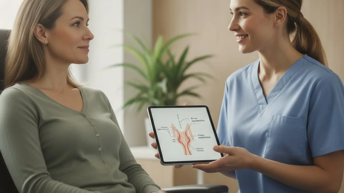
Fine Needle Aspiration: The Thin-Needle Biopsy
The report that comes back describes what those cells look like under a microscope. A clinician reads that description next to your examination and your imaging, then decides what happens next.
What is a thin-needle biopsy, and how does it differ from other biopsies?
It is a procedure in which a clinician passes a very thin needle into a lump or an organ, moves it through the tissue a few times, and draws loose cells into the needle for a laboratory to examine. Nothing is cut, nothing is stitched, and the material that reaches the laboratory is a scattering of cells rather than a piece of tissue.
That is why the laboratory discipline involved is cytology. You will often see the phrase fine needle aspiration cytology on a request form or a report, and it simply means the specialist is reading individual cells and small clusters of cells rather than a slice of tissue with its original structure intact.
The comparison most people want is with the other needle procedure. A core needle biopsy uses a wider, hollow needle to remove a small cylinder of tissue, so the pathologist can see not only what the cells look like but how they are arranged within the tissue and whether they have pushed past their normal boundary. A surgical biopsy goes further again and removes part or all of the lump in a procedure room or operating room.
All three are biopsies. They differ in how much material they collect, how invasive they are, and what question the laboratory can answer from what arrives. Research suggests that thin-needle sampling is central to the assessment of thyroid nodules, where it is used to sort nodules that need an operation from those that can be followed 1. A clinician chooses between the three based on the site, the imaging appearance and what needs to be answered.
Why would a clinician suggest sampling a lump this way?
Because imaging can describe a lump's size, shape, borders and blood flow, but it cannot describe the cells inside it. A scan can say a nodule looks concerning or looks unremarkable. Only a sample puts cells under a microscope.
The practical appeal is the balance of information against intrusion. The procedure is usually done in a clinic room rather than an operating room, it does not require an incision, and the equipment is a syringe, a fine needle and often an ultrasound probe. When a lump is fluid filled, fluid can be drawn off at the same sitting and sent for examination.
The result then feeds a decision. Depending on what the cells look like, the conversation that follows may be about watching the lump with repeat imaging, repeating the sample, adding further laboratory testing on the material already collected, or referral to a surgeon. Research suggests that needle sampling is the pivotal test for deciding which thyroid nodules are referred for an operation and which are monitored over time 1.
It is worth being clear about the limit as well. The sample answers a question about cells in the area the needle reached. It does not answer every question about the lump, and a clinician decides whether the answer it gives is enough or whether a larger sample is needed.
How is the procedure done, and how do you prepare?
The appointment is short and follows a predictable sequence. You lie or sit so the lump is easy to reach, the skin over it is cleaned, and the clinician locates the target, often with an ultrasound probe held beside the needle so the tip can be watched as it moves.
For a neck lump, the head is usually tilted back on a pillow to bring the thyroid forward. You may be asked not to swallow, cough or talk while the needle is in place, because the thyroid rides up and down with swallowing. The clinician then makes several brief passes, each lasting a few seconds, moving the needle within the lump to pick up cells from more than one spot. Several passes from slightly different angles are normal and are about getting enough material, not about anything having gone wrong.
A local numbing medicine is sometimes used for the skin, and sometimes not, because the numbing injection itself involves a needle. That choice belongs to the clinician performing the procedure, and it is reasonable to ask in advance.
Preparation is usually minimal, and the instructions that matter are the ones the clinic performing the procedure gives you. Tell the team about any bleeding problems and about everything you take, including things bought without a prescription. Some medicines affect bleeding, so ask your prescriber well before the appointment and do not stop anything on your own. Afterwards, pressure is held over the site and a small dressing is applied.
Does a thyroid needle test hurt?
Most descriptions centre on pressure and a short sting rather than deep pain, but the experience varies from person to person and no one can predict yours in advance. The needle is fine, each pass is brief, and the uncomfortable part is usually the moment the needle crosses the skin.
A neck procedure has its own small discomforts that are not pain exactly. Lying with the head tilted back strains the neck for people with stiff necks or back problems, and the instruction not to swallow can feel harder than it sounds. Asking for a pillow under the shoulders or a short pause between passes is reasonable.
Soreness at the site afterwards is commonly described, and it can feel like a bruise when you turn your head. How long that lasts differs between people, so rather than expecting a set timeline, note what you feel and tell the clinic if soreness increases rather than settles, or if swelling grows.
If pain during the procedure is more than you expected, say so while it is happening. The clinician can stop, add numbing or change the approach. Pain is information, not something to sit through quietly.
What happens to the sample after it leaves the room?
The cells are spread onto glass slides, fixed and stained, and in many laboratories some of the material is also placed in liquid so it can be spun down and processed into a small block for further testing. A cytopathologist then examines the slides.
The first judgement is whether there is enough well preserved material to interpret at all. Cells that are too few, obscured by blood, or damaged in handling limit what can be said, which is why adequacy is assessed before anything else. Some centres have a specialist assess adequacy while you are still in the room so that another pass can be taken immediately if needed.
If the appearance raises a specific question, extra testing can often be run on material already collected. Stains that highlight particular proteins help separate cell types that look similar, and samples from lymph nodes can be sent for cell-by-cell marker testing on the same aspirate. For thyroid samples that fall into an uncertain category, additional laboratory testing on the collected cells is one of the options a clinician may consider 2.
The report that results is descriptive. It sets out what the cells look like, places them in a reporting category where a standard system exists, and often adds a comment on what the clinician may wish to consider. Interpretation of what that means for you belongs with the clinician who ordered it.
How is a thyroid nodule evaluated with a needle sample?
The nodule is first assessed on ultrasound, and the features seen there, together with its size, guide whether sampling is offered at all. Thyroid nodules are common, and not every one is sampled.
When sampling is offered, it is usually done with ultrasound guidance so the needle tip can be watched entering the part of the nodule most worth sampling, such as a solid area within a partly fluid-filled nodule. Research suggests that needle sampling, read alongside ultrasound features, is the main tool for working out which thyroid nodules need surgical referral 1.
The resulting report uses a standard set of categories rather than free text, so that clinicians in different places read the same words the same way. Standardised reporting, together with laboratory testing that can be added to uncertain samples, is described as the current shape of thyroid cytopathology practice 2.
Two things are worth holding onto. First, a needle sample describes a nodule, not the whole gland, and a gland can contain several nodules that would not all give the same answer. Second, the sample says nothing about whether the gland is making too much or too little hormone, which is a blood-work question. A result outside the printed range on any thyroid blood test is worth discussing with a clinician, who decides what it reflects.
What does needle sampling add in a breast workup?
It supplies the cell-level part of a three-part assessment, alongside the clinical examination and the imaging. A breast lump is rarely judged on one of those three alone, and the strength of the approach comes from reading them together.
Needle sampling of the breast is associated with recognised interpretive pitfalls, which is one reason reports are read next to the examination findings and the images rather than in isolation 3. Certain lesions can look more worrying on cells alone than they turn out to be, and others can look deceptively unremarkable, so a cytopathologist weighs several features rather than one.
The reporting of breast cytology has been refined over time, with clearer criteria for placing a sample into a category rather than leaving it as an impression 4. Practice also varies by centre and by lesion: many breast workups now use a core needle biopsy when tissue structure matters, while thin-needle sampling continues to be used for fluid-filled lumps, for sampling nearby lymph nodes and in settings where a quick cell answer is useful.
Breast lumps in men are assessed with the same tools. Research suggests that thin-needle sampling has diagnostic value in male breast lesions 5. Any new breast lump, in anyone, is worth bringing to a clinician rather than watching at home.
Can a swollen lymph node or another site be sampled the same way?
Yes, and lymph nodes are one of the places where the approach is used most. A node that can be felt in the neck, armpit or groin can often be sampled in a clinic room, with ultrasound guidance if it sits deeper.
What the sample can answer depends on the question. Cell appearance alone can be enough when the question is whether cells from a cancer elsewhere have travelled to the node. When the question involves the lymph system itself, marker testing is added to the aspirate. Evidence suggests that combining needle sampling with cell-by-cell marker testing extends what can be assessed from a lymph node aspirate, while full classification may still call for a tissue sample 6.
The approach is not limited to the thyroid, breast and lymph nodes. Salivary gland lumps, superficial soft tissue lumps and some deeper masses reached with imaging guidance are sampled the same way. Studies indicate that thin-needle cytology also has a role in assessing gynaecologic tumours, including masses that recur or spread beyond their original site 7.
What stays constant across sites is the trade-off. A fine needle gives cells quickly with little intrusion. When the answer needs tissue structure, a larger sample follows, and the clinician decides when that point has been reached.
What do the result categories on the report mean?
They are standard descriptive labels for what the cytopathologist saw, not verdicts about you. Thyroid reporting in particular uses a named set of categories so that the same appearance is described the same way in different laboratories, and the categories are usually written out in words on the report.
| Category as written on a thyroid report | What it describes | What is usually discussed next |
|---|---|---|
| Nondiagnostic or unsatisfactory | Too few usable cells, or cells obscured by blood or poor preservation | Repeating the sample, often with ultrasound guidance |
| Benign | Cells with the appearance of a noncancerous nodule | Follow-up imaging on a schedule the clinician sets |
| Atypia of undetermined significance | Some cells look unusual, but not enough to place the sample in a firmer category | Repeat sampling, further laboratory testing, or closer follow-up |
| Follicular neoplasm or suspicious for a follicular neoplasm | A cell pattern that cytology alone cannot separate from a lookalike | Surgical opinion or further laboratory testing on the sample |
| Suspicious for malignancy | Features that point towards cancer without meeting full criteria | Surgical opinion |
| Malignant | Features of cancer in the cells examined | Referral and staging decisions made with a specialist team |
Breast and lymph node samples use their own reporting frameworks with similar logic, moving from inadequate, through uncertain, to definite. Whatever the site, a category describes the cells on the slide. It does not announce a condition, and the clinician who ordered the test decides what it reflects for you and what follows.
Why does a result come back nondiagnostic or atypical, and what happens then?
Because a fine needle collects a small number of cells from a small part of a lump, and sometimes those cells either do not arrive in usable condition or do not look clearly like anything. Neither outcome means the procedure was done badly.
Several things influence the yield. Fluid-filled nodules can give mostly fluid and few cells. Very firm or scarred lumps release cells reluctantly. Bleeding into the sample obscures the slide. Deeper or small targets are harder to hit without ultrasound guidance, and how the slides are prepared affects how well the cells survive the journey.
Atypical results are a different problem. Here the cells arrived but sat between categories. Studies indicate that the definition of atypia in thyroid cytology has been revised over successive versions of the reporting system as criteria were tightened 8. A structured approach to atypical thyroid findings, including recognising appearances that mimic more worrying ones, is described in the cytopathology literature 9.
What happens next is usually one of three things. The sample is repeated, sometimes after an interval and usually with ultrasound guidance. Further laboratory testing is run on material already collected. Or the lump is followed with imaging on a defined schedule. A repeat is a normal step in the pathway, not a setback, and the choice between these belongs to the clinician who knows the whole picture.
How does this compare with a core needle biopsy or surgery?
The three sit on a ladder, from least material and least intrusion to most of both. Choosing between them is about what question needs answering, not about which is better.
| Feature | Thin-needle sampling | Core needle biopsy | Surgical biopsy |
|---|---|---|---|
| What is collected | Loose cells and small cell clusters | A small cylinder of intact tissue | Part or all of the lump |
| Instrument | A fine needle on a syringe | A wider hollow needle, often spring loaded | A scalpel, in a procedure or operating room |
| Numbing | Skin numbing sometimes used, at the clinician's discretion | Local numbing used | Local or general anaesthesia |
| What the laboratory can assess | Cell appearance, with added stains or marker testing when ordered | Cell appearance plus how cells are arranged in the tissue | The whole lesion and its edges |
| Stitches | None | Usually none | Yes |
| Common uses | Thyroid nodules, lymph nodes, fluid-filled lumps, salivary lumps | Many breast and soft tissue lesions where structure matters | When needle sampling leaves the question open |
The ladder is often climbed rather than jumped. A thin-needle sample that answers the question ends the process. One that comes back uncertain or inadequate leads to a repeat, a core sample or a surgical opinion. Activity and aftercare following any of them are set by the clinician who performs the procedure.
What are the risks, and what should prompt urgent attention?
The common complaints are local and mild: soreness, bruising, a small amount of bleeding at the puncture site, and swelling over the area. Pressure after the needle is withdrawn is used to limit bruising.
Less common problems include bleeding into the lump itself, which can make it feel larger for a time, and infection at the site. Neck procedures carry a small risk of swelling in a confined space, and procedures near the lung wall carry a small risk of air leaking into the chest, which is why clinicians choose the approach angle carefully and use imaging guidance.
Call 911 or go to the emergency department for trouble breathing, for swelling in the neck that is growing quickly, for difficulty swallowing that comes on with that swelling, for bleeding that does not stop with firm pressure, or for chest pain or fainting. Do not wait to see whether these settle.
Contact the clinic that performed the procedure promptly for a fever, for spreading redness or warmth around the site, for pain that increases over days rather than easing, or for any new change at the site you were not told to expect. Describing what you notice in plain terms is enough; the clinician decides what it reflects.
Where does TSH fit into a thyroid nodule workup?
Blood work answers a different question from a needle sample. TSH is the pituitary signal laboratories use to gauge how hard the thyroid is being pushed, and it is usually checked when a nodule is found, because the result can change which test is ordered next.
FitScript's optimal range for TSH is 0.5 to 2 mIU/L, which sits alongside your laboratory's own range as printed on your lab report. FitScript's optimal range is an optimisation target, not a diagnostic cut-off. A value outside the range printed on your report is worth discussing with a clinician, who decides what it reflects.
The deeper explanation of what this marker measures, how it is tested and how to read a result lives on its own page.
Which other tests are usually ordered alongside?
Imaging comes first in almost every pathway, because it decides whether a needle is used at all and where it goes. For a neck lump that means ultrasound of the thyroid and the surrounding lymph nodes. For a breast lump it may mean mammography, ultrasound or both, read with the clinical examination.
Blood work runs in parallel rather than in competition. Thyroid function markers describe how the gland is working. General markers such as a full blood count give context when a lymph node is the concern. None of these describe what cells inside a lump look like, which is exactly the gap a needle sample fills.
A membership like FitScript, with 150+ biomarkers tested per year and interpretation from Atlas, its AI health advisor, covers the blood-work side of that picture and tracks it over time. It does not replace imaging or a procedure a clinician performs, and nothing in a panel substitutes for sampling a lump that a clinician wants sampled.
The practical move is to keep the pieces together. Bring your imaging reports, your recent blood work and the cytology report to the same appointment, so the clinician reading them is not working from one fragment. Knowing how your own markers have moved over the years makes that conversation shorter and sharper.
When should you talk to a clinician?
About any new lump, any lump that is growing, and any lump you have been told to watch that has changed. That is true whether it is in the neck, the breast, the armpit, the groin or anywhere else, and it is true for men as well as women.
Bring it forward sooner when a neck lump comes with a hoarse voice that is not settling or with difficulty swallowing, when a lump is hard and fixed in place rather than moving under the skin, or when a swollen node comes with unexplained fever, drenching night sweats or weight loss you did not intend. These are reasons to be seen, not reasons to panic, and the clinician decides what they reflect.
Also book a conversation about results you do not understand. A report that reads nondiagnostic, atypical or suspicious deserves an explanation in plain language, including what the next step is and when it happens. Screening decisions, and whether any further testing is right for you, are a talk-with-your-clinician matter rather than something to settle from an article.
This article is educational and is not medical advice. Discuss your results and any treatment with a clinician or licensed provider, and remember that treatment decisions belong to a licensed provider who has reviewed your full situation.
Key takeaways
- Fine needle aspiration collects loose cells through a very thin needle, so the laboratory reads cytology rather than tissue structure.
- It is one of three biopsy options, alongside core needle sampling and surgical removal, and the clinician picks the one that answers the question at hand.
- Thyroid, breast and lymph node lumps are the most familiar targets, and ultrasound guidance is often used to place the needle.
- Reports use standard category names rather than verdicts; the clinician who ordered the test decides what a category reflects for you.
- Nondiagnostic and atypical results are recognised parts of the pathway, and a repeat sample or further laboratory testing is a normal next step.
- Blood markers such as TSH describe how the thyroid is functioning; they do not describe the cells inside a nodule, and the two tests answer different questions.
- Call 911 or go to the emergency department for trouble breathing, quickly growing neck swelling, difficulty swallowing with that swelling, uncontrolled bleeding, chest pain or fainting.
References
- Fine Needle Aspiration in the Investigation of Thyroid Nodules. Deutsches Arzteblatt international, 2016.
- Thyroid Fine-Needle Aspiration: The Current and Future Landscape of Cytopathology. Surgical pathology clinics, 2024.
- Overcoming Pitfalls in Breast Fine-Needle Aspiration Cytology: A Practical Review. Acta cytologica, 2024.
- Fine needle aspiration (FNA) cytology of breast: refining the diagnosis. Cytopathology, 1998.
- Diagnostic Value of Fine-Needle Aspiration in Male Breast Lesions. Acta cytologica, 2019.
- Fine-needle aspiration cytology in the diagnosis of lymphoma. The next step. American journal of clinical pathology, 2000.
- Role of Fine Needle Aspiration Cytology in the Diagnosis of Gynecologic Tumors. Acta cytologica, 2023.
- The evolution of 'atypia' in thyroid fine-needle aspiration specimens. Diagnostic cytopathology, 2022.
- Pitfalls in Thyroid Fine-Needle Aspiration Cytopathology: An Approach to Atypical Findings. Acta cytologica, 2024.
Common questions
Is fine needle aspiration the same thing as a biopsy?
It is a type of biopsy. The difference is how much material is collected: a fine needle draws out loose cells, while a core needle biopsy removes a small cylinder of tissue and a surgical biopsy removes part or all of the lump. All three send material to a laboratory, but each lets the pathologist assess a different amount of detail.
Does a thyroid needle test hurt?
People usually describe pressure and a brief sting rather than deep pain, though the experience differs from person to person. Lying with the neck extended and being asked not to swallow can feel more awkward than the needle itself. Soreness afterwards varies, so tell the clinic if it increases instead of easing.
How long does the procedure take?
The needle part is brief, often only a few seconds per pass, with several passes taken to collect enough cells. The appointment itself is longer because of positioning, cleaning the skin, ultrasound setup and holding pressure afterwards. Ask the clinic performing it what to expect for their setup.
Do I need to fast or stop anything beforehand?
Follow the instructions the clinic performing the procedure gives you, since preparation varies by site and by setting. Tell the team about any bleeding problems and about everything you take, including items bought without a prescription. Some medicines affect bleeding, so ask your prescriber in advance and do not stop anything on your own.
What does a nondiagnostic result mean?
It means the laboratory did not receive enough usable cells to interpret, often because the sample was mostly fluid, obscured by blood or poorly preserved. It is not a finding about your health and it does not mean anything was done wrong. The usual next step is repeating the sample, often with ultrasound guidance, and your clinician decides on the timing.
Why would a result be called atypical?
Atypical means the cells arrived in usable condition but did not look clearly like one category or another. It is an honest description of uncertainty rather than a conclusion. What follows is usually a repeat sample, further laboratory testing on the material already collected, or closer follow-up imaging, and the clinician who ordered the test decides which.
Can a swollen lymph node be sampled this way?
Yes. Nodes that can be felt in the neck, armpit or groin are often sampled in a clinic room, and deeper nodes can be reached with ultrasound guidance. Extra marker testing can be run on the same sample when needed, and a clinician decides whether a larger tissue sample is required afterwards.
What should I watch for after the procedure?
Call 911 or go to the emergency department for trouble breathing, neck swelling that is growing quickly, difficulty swallowing with that swelling, bleeding that does not stop with firm pressure, chest pain or fainting. Contact the clinic that performed the procedure promptly for fever, spreading redness around the site, or pain that increases over days. Describe what you notice and let the clinician decide what it reflects.