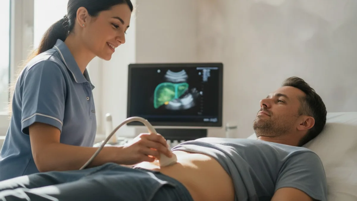
Liver Ultrasound: Fatty Liver, Stiffness and Results
Yellow eyes with confusion, vomiting blood, or a belly that swells quickly need emergency care. Call 911 rather than waiting for a scan appointment.
What does the scan actually look at?
It looks at four things at once, the size and outline of the liver, how bright and how even the tissue appears, the bile ducts and gallbladder sitting against it, and the direction and speed of blood moving through the portal and hepatic veins.
The physics is straightforward. A handheld transducer sends high frequency sound pulses into the abdomen and listens for the echoes that return. Tissue that reflects more sound looks brighter on the grayscale image, tissue that reflects less looks darker, and the machine times each returning echo to work out how deep it came from. Because the picture refreshes many times a second, the sonographer watches the liver slide under the ribs as you breathe rather than capturing one frozen slice. A Doppler setting adds blood flow on top of the grey picture, showing whether blood is travelling toward the liver as it normally does.
Two practical features matter. There is no radiation, so the scan can be repeated as often as a clinician thinks useful, and it is done in real time, so the person holding the probe can chase an unclear area from a different angle.
What the scan cannot do is measure function. It does not tell you how well the liver is clearing bilirubin, building albumin or handling the chemistry of a meal. It describes appearance and flow. Blood markers describe activity and capacity. Neither one replaces the other, and a clinician reads them side by side.
Why would a clinician order one?
Most liver scans are ordered because something else raised a question first, usually liver enzymes sitting outside the standard lab reference range on a routine panel, discomfort or fullness under the right ribcage, or a metabolic picture that makes fat in the liver more likely, such as rising fasting glucose, high triglycerides and weight carried around the middle.
It is also the usual first imaging test when fat in the liver is suspected, because it is widely available, carries no radiation and costs far less than cross sectional imaging. Research suggests ultrasound based methods are the most commonly used first line approach for assessing fat in the liver 1.
The other common reason is follow up. Once a report has described fatty change, a clinician may want to see whether the picture has shifted after several months of changes to food, movement, alcohol intake and body composition.
It is worth being clear about what an order means. A request for a scan is not a statement that something is wrong. It is a way of converting one odd number into a picture, so that the next conversation is based on more than a single value. A raised enzyme on its own is an observation. What it may reflect, and what should happen next, is for a clinician to work out with the full set of results in front of them.
What does a bright or echogenic liver mean?
A bright, or echogenic, liver means the tissue is reflecting more sound than expected, and the most common reason for that in adults is fat stored inside liver cells. Fat droplets create many small boundaries inside the tissue, and each boundary sends part of the sound pulse back toward the probe, so the whole organ lights up.
Radiologists usually judge brightness by comparison rather than in isolation. The liver is held up against the right kidney in the same image, since the two normally look similar in brightness. A liver that is clearly brighter than the kidney next to it, with vessel walls that lose their crisp outline and a diaphragm that becomes harder to see at depth, is the classic described pattern. These grayscale features are the ones that have long been used to identify fat in the liver 1.
Reports often grade this as mild, moderate or severe. Mild usually means a slight increase in brightness with vessel borders still visible. Moderate means those borders start to blur. Severe means the sound beam is absorbed so strongly on the way through that the deep part of the liver and the diaphragm become difficult to see at all.
Here is the honest limit of the phrase. Increased echogenicity is a description of how tissue looks on a screen. It is strongly suggestive of fat, it is not a diagnosis by itself, and a licensed provider is the one who decides what the finding means for you and whether anything further is needed.
Can the scan miss fat in the liver?
Yes, particularly when the amount of fat is small. A meta-analysis of ultrasonography for detecting fatty liver found good accuracy for moderate to severe fatty change compared with biopsy 2, and the same work showed that performance falls away when only a small proportion of liver cells contain fat 2.
That has a practical consequence. A report that says "normal hepatic echotexture" makes a lot of fat unlikely, but it does not rule out early fatty change. This is one of the reasons bloodwork and the scan are read together rather than one being used to dismiss the other.
Two other limits sit behind the same problem. Grayscale brightness is a visual judgement, so two readers can grade the same liver slightly differently, and a thick abdominal wall weakens the sound beam before it ever reaches the liver, which makes the deep tissue look darker and the whole assessment harder.
Newer ultrasound measurements try to put a number on fat rather than an adjective, by measuring how quickly sound energy fades as it passes through the tissue. International guidance has been published on how these fat quantification measurements should be acquired and reported 3, and that guidance notes that values are not freely interchangeable between machines from different manufacturers 3. If your clinic offers one, ask which measurement was used, so that a future scan can be compared like for like.
How does elastography measure stiffness?
Elastography measures stiffness by watching how fast a mechanical wave travels sideways through liver tissue. The probe sends a brief push into the liver, that push generates a shear wave that ripples outward, and the machine tracks how quickly the ripple moves. Stiffer tissue carries the wave faster, softer tissue carries it slower, and the speed is converted into a stiffness value.
It happens inside the same appointment on many modern machines, takes a few extra moments, and feels like nothing more than the probe resting between two ribs. The main things asked of you are to lie still, breathe quietly and hold a relaxed breath for each measurement.
How the measurement is taken changes the answer, which is why consensus documents exist. A consensus statement from a radiology society sets out standardised acquisition for liver stiffness, including probe position, patient breathing and the number of measurements collected 4. International guidance covering liver elastography similarly standardises how measurements should be acquired and reported so that results travel between clinics 5. Reviews of the technique describe the same core principle, that shear wave speed through the tissue is used as a surrogate for tissue stiffness 6.
One detail worth knowing before you go. Measurement is usually requested in a fasted state, because a recent meal increases blood flow to the liver and can push the reading up 4.
What can a stiffness reading suggest about scarring?
A higher stiffness value suggests more fibrous tissue in the liver, because scar tissue is physically stiffer than healthy liver. Liver stiffness measured by ultrasound or magnetic resonance elastography is associated with the amount of fibrosis present in the tissue 7.
In people with fatty liver disease specifically, studies suggest that ultrasound elastography may help separate those with more advanced fibrosis from those with little or none 8. A meta-analysis comparing routine laboratory tests, ultrasound elastography and magnetic resonance elastography for detecting fibrosis in nonalcoholic fatty liver disease found magnetic resonance elastography performed best of the approaches compared 9.
Two cautions keep this honest. First, a stiffness number is an estimate of a tissue property, not a tissue sample, and the thresholds used to interpret it differ by machine, by technique and by the underlying reason for liver disease, which is why your report should be read by the clinician who ordered it rather than against a number found online. Second, stiffness is not only about scar. Inflammation, congestion of blood in the liver and a recent meal can all raise the reading, and consensus guidance specifically flags these as conditions that need to be accounted for when interpreting a value 4.
So the sensible way to read a stiffness result is as one input. A low value is reassuring about advanced scarring. A raised value is a reason for a clinician to look harder, repeat the measurement under better conditions, or order something else, not a conclusion on its own.
Where does ALT fit into the picture?
ALT is the blood side of the same question. The scan describes structure, how bright the tissue looks and how stiff it measures. ALT is an enzyme concentrated inside liver cells, and more of it appears in the blood when those cells are turning over faster, so it reports activity rather than appearance. The two often move together, and sometimes they do not.
FitScript publishes an optimal range for ALT that sits tighter than the standard lab reference range, and that optimal range is an optimization target rather than a diagnostic cut off. Standard reference ranges themselves vary by lab, by age and by sex, so always read your value against the range printed on your own report. The ALT guide linked above covers what the marker measures, how it is tested and how to interpret a single result.
Which blood tests belong alongside the scan?
The scan is most useful next to a liver panel plus the metabolic markers that explain why fat accumulates in the liver in the first place. A clinician decides which of these to order, and the list below is about what each one adds rather than a shopping list to request.
| Test | What it adds next to the scan |
|---|---|
| AST | A second enzyme from liver cells, which clinicians read next to ALT rather than alone |
| ALP and GGT | Point more toward bile flow and the biliary system than toward liver cell turnover |
| Bilirubin | Reflects how well pigment from red cell breakdown is being cleared |
| Albumin | A protein the liver makes, so it speaks to synthetic capacity over time |
| Platelet count | Used inside several routine blood based fibrosis index scores |
| Fasting glucose, insulin and HbA1c | Describe the metabolic pressure that drives fat storage in the liver |
| Triglycerides and HDL | The lipid pattern that commonly travels with fatty change |
| Ferritin and iron markers | Iron handling influences both lab values and tissue appearance |
| Viral hepatitis screening | Ordered at a clinician's discretion to look for other causes of abnormal enzymes |
This is where regular testing earns its place. FitScript members have 150+ biomarkers tested per year, with results interpreted by Atlas, the AI health advisor, so a single scan sits inside a trend rather than standing alone.
How do I read the wording on my report?
Read it as description first, then meaning, then next step, because radiology reports are written to describe what the sonographer saw, not to name a condition. The table below translates the phrases that appear most often.
| Report phrase | What it describes | What it does not establish |
|---|---|---|
| Increased echogenicity | The tissue reflects more sound than expected | The cause of the brightness |
| Hepatic steatosis, mild or moderate | A graded impression of fat in the liver | How much inflammation or scarring is present |
| Normal echotexture | The tissue looks even and of expected brightness | That small amounts of fat are absent |
| Hepatomegaly | The liver measures larger than expected | Why it is enlarged |
| Smooth contour | The surface outline looks regular | That the tissue is healthy throughout |
| Portal vein patent with normal direction of flow | Blood is moving into the liver as expected | Anything about tissue stiffness |
| No biliary dilatation | The bile ducts are not widened | Anything about liver cell turnover |
| Limited scan due to body habitus or bowel gas | Image quality was reduced | That the liver is normal or abnormal |
That last row matters more than people expect. If the report says the view was limited, the scan answered less than it looks like it did, and the clinician who ordered it may want to repeat it or choose a different test.
How is the scan done, and how should I prepare?
You lie on your back or turned slightly onto your left side, a warm gel goes on the skin over the right ribcage, and the sonographer presses a probe between and under the ribs while asking you to take and hold a breath. The breath hold is not ceremony. It drops the diaphragm and pushes the liver down out of the rib shadows, which is the only way to see the upper segments clearly.
Most clinics ask for a period of fasting beforehand, which empties the gallbladder so its wall can be seen properly and reduces bowel gas sitting in the way. Fasting also matters for stiffness measurement, since a recent meal raises liver blood flow and can raise the reading 4. Scan preparations vary between clinics, so follow the instructions the imaging department gives you rather than a general rule, and confirm how long the appointment is expected to take when you book.
There are no needles, no radiation and nothing you need to swallow for a standard abdominal scan. Wear something that lets you expose the upper abdomen easily. The firm pressure under the ribs can feel odd, and occasionally tender if the area is already sore, so say so and the sonographer will adjust.
Results usually go to the clinician who ordered the scan rather than being discussed in the room, since the sonographer captures images and a radiologist writes the report. Ask when and how you will receive it, and book the follow up conversation at the same time.
What can throw a result off?
Several everyday things change what the scan shows, which is why one measurement is weaker than a trend. The biggest is body habitus. A thicker abdominal wall absorbs sound before it reaches the liver, dimming the deep tissue and making both brightness grading and stiffness measurement harder and less reliable.
Technique is the second. Grayscale assessment of fat depends on gain settings, probe choice and the reader's judgement, which is part of why numerical fat quantification methods were developed and why guidance standardises how they should be performed and reported 3.
For stiffness, the list of influences is well described. Food in the last few hours, congestion of blood in the liver, active inflammation and even breathing against a held breath can all shift the number, and consensus guidance builds protocol rules around exactly these factors 4. International guidance adds that measurements from different systems and techniques are not automatically interchangeable, so comparing this year's value with last year's from another machine can mislead 5.
Some medicines can also affect liver enzymes and the broader picture a clinician is reading alongside the scan. Do not stop or change anything on your own, and ask your prescriber whether anything you take is relevant before the appointment.
The workaround is simple. Where possible, repeat scans at the same clinic, on the same type of equipment, under the same preparation conditions.
What besides fat can make a liver look bright or stiff?
Fat is the most common explanation for a bright liver in adults, but it is not the only one, and stiffness has an even longer list of contributors. Fibrous tissue laid down over time changes both how the liver reflects sound and how fast a shear wave travels through it, and stiffness measured by elastography is associated with the degree of fibrosis in the tissue 7.
Inflammation is the second contributor. Swollen, inflamed liver tissue measures stiffer than quiet tissue at the same amount of scarring, which is one reason consensus guidance asks for interpretation in the clinical context rather than against a single cut off 4.
Congestion is the third. When blood backs up into the liver from the right side of the heart, the organ becomes engorged and stiffer on measurement, which is listed among the conditions that need to be considered when a stiffness value is read 4.
Iron and other stored substances change how tissue behaves on imaging too.
The practical takeaway is to resist pinning a cause on a picture. A bright or stiff liver is a finding that narrows the list of possibilities. Sorting that list is a clinical job that uses your history, your alcohol intake, your metabolic markers and sometimes further testing.
How does it compare with other ways of checking fat and scarring?
Ultrasound is the accessible first look, magnetic resonance based imaging is the most accurate non invasive option, and biopsy remains the reference that the others are measured against.
| Approach | What it measures | Strengths | Limits |
|---|---|---|---|
| Grayscale ultrasound | Brightness and texture of the tissue | Fast, no radiation, widely available, inexpensive | Graded by eye, less sensitive when fat is minimal |
| Ultrasound fat quantification | How quickly sound energy fades through the liver | Produces a number that can be tracked over time | Values differ between manufacturers and are harder in larger bodies |
| Ultrasound elastography | Shear wave speed as a measure of stiffness | Done in the same visit, protocol driven, repeatable | Affected by food, inflammation and congestion |
| Magnetic resonance based imaging | Fat fraction and tissue stiffness across the whole organ | Highest non invasive accuracy in the comparison below | Cost, access and scan time |
| Blood based fibrosis index scores | A calculated estimate from routine lab values | Cheap, repeatable, no appointment needed | Indirect, with results that often land in an indeterminate band |
| Liver biopsy | Tissue under a microscope | Direct assessment of fat, inflammation and fibrosis | Invasive, and samples a very small part of the organ |
The comparison between these has been measured directly. A meta-analysis in people with nonalcoholic fatty liver disease compared laboratory tests, ultrasound elastography and magnetic resonance elastography for detecting fibrosis and found magnetic resonance elastography the most accurate of the three 9. Research also suggests ultrasound elastography performs well enough to be useful in routine assessment of this group 8, which is why the cheaper, faster test is usually the one done first and the expensive one is reserved for when the answer needs to be firmer.
What if the scan and the bloodwork disagree?
Disagreement between imaging and labs is common, and it is information rather than a contradiction. Each of the three patterns below has a sensible next step, and in every case the next step belongs to the clinician holding your full history.
A normal looking scan with raised enzymes
What the scan shows is that large amounts of fat are unlikely, since detection falls off when only a small proportion of cells are involved 2. What raised enzymes may reflect is anything that increases liver cell turnover, including early fatty change below the threshold the scan can see, recent intense exercise, alcohol intake, or other causes a clinician would consider. The useful next step is a repeat panel after a period without alcohol and without unusually heavy training, plus a conversation about everything you take, including things bought without a prescription.
A bright liver with normal enzymes
This combination is frequent and often surprises people. Fat can be present in the tissue while enzymes sit inside the standard reference range, because storing fat and damaging cells are not the same process. The reasonable next step is to look at the metabolic markers around it, fasting glucose and insulin, HbA1c, triglycerides and waist measurement, and to decide with a clinician whether stiffness assessment adds anything.
A raised stiffness value with everything else unremarkable
Treat this as a reading to confirm rather than accept. Check whether you were fasted, whether the measurement quality indicators were acceptable and whether anything that raises blood flow or inflammation was present on the day, since consensus guidance identifies these as factors that influence the value 4. A repeat under proper conditions, on the same equipment, is often the most informative move, and the clinician who ordered it decides whether a different test is warranted.
When is it worth talking to a clinician?
Always, if a report uses a word you cannot translate, and promptly if a result sits outside the standard reference range or a stiffness value is flagged on your report. Do not ignore an abnormal result because you feel well, and do not act on one in isolation either. A suboptimal value is a reason for a conversation, not a conclusion.
Bring four things to that conversation. Your current report and any previous scan, your recent lab panel, an honest account of alcohol intake, and a full list of everything you take, including supplements, because some medicines affect liver markers and your prescriber is the person to ask about yours. Treatment decisions belong to a licensed provider who has evaluated you.
Call 911 or go to the emergency department, rather than waiting for an appointment, for yellowing of the eyes or skin together with confusion or drowsiness, for vomiting blood or passing black tarry stools, or for a belly that swells quickly with pain. Yellowing of the skin or eyes on its own, without those features, still warrants being seen quickly.
This page is educational and not medical advice.
If you want to go deeper on the two things that sit either side of this scan, what do liver function tests show explains how the standard blood panel is read, and yellow skin causes walks through why skin and eyes can take on a yellow tone and which of those reasons need attention sooner.
Key takeaways
- The scan describes structure, brightness, size and blood flow. It does not measure how well the liver is working.
- A bright, or echogenic, liver usually suggests fat stored in liver cells, and remains a description rather than a diagnosis.
- Detection of moderate to severe fatty change is good, while smaller amounts are often missed 2.
- Elastography estimates stiffness from shear wave speed, and stiffness is associated with the amount of fibrosis in the tissue 7.
- Food, inflammation, congestion and equipment differences all move stiffness values, so repeat under the same conditions where you can 4.
- Read the scan next to a liver panel and metabolic markers, and take an abnormal result to a clinician rather than to a search bar.
References
- Ultrasound-based techniques for the diagnosis of liver steatosis. World Journal of Gastroenterology, 2019.
- Diagnostic accuracy and reliability of ultrasonography for the detection of fatty liver: a meta-analysis. Hepatology, 2011.
- WFUMB Guidelines/Guidance on Liver Multiparametric Ultrasound. Part 2: Guidance on Liver Fat Quantification. Ultrasound in Medicine & Biology, 2024.
- Update to the Society of Radiologists in Ultrasound Liver Elastography Consensus Statement. Radiology, 2020.
- WFUMB Guideline/Guidance on Liver Multiparametric Ultrasound: Part 1. Update to 2018 Guidelines on Liver Ultrasound Elastography. Ultrasound in Medicine & Biology, 2024.
- Ultrasound elastography: liver. Abdominal Imaging, 2015.
- Liver fibrosis assessment: MR and US elastography. Abdominal Radiology (New York), 2022.
- Advances in ultrasound elastography for nonalcoholic fatty liver disease. Journal of Medical Ultrasonics, 2020.
- Comparison of laboratory tests, ultrasound, or magnetic resonance elastography to detect fibrosis in patients with nonalcoholic fatty liver disease: A meta-analysis. Hepatology, 2017.
Common questions
Do I need to fast before a liver ultrasound?
Most clinics ask you to fast beforehand, because an empty gallbladder is easier to see and there is less bowel gas blocking the view. Fasting also matters if a stiffness measurement is being taken, since a recent meal can push the reading up. Follow the exact preparation instructions your imaging department gives you, as they vary between clinics.
Does the scan hurt, and does it use radiation?
No radiation is involved and there are no needles for a standard abdominal scan. You will feel firm pressure from the probe under the right ribs and some cool gel on the skin, which can be mildly uncomfortable if the area is already sore. Tell the sonographer if it is painful and they will change the angle or pressure.
My report says my liver is bright or echogenic. What does that mean?
It means the tissue reflected more sound than expected, which in adults most often points toward fat stored inside liver cells. It is a description of appearance, not a diagnosis, and several other things can produce the same look. Take the report to the clinician who ordered it so it can be read alongside your bloodwork and history.
Can a scan miss fat in the liver?
Yes, particularly when the amount of fat is small. A report describing normal echotexture makes a large amount of fat unlikely, but it does not rule out early fatty change. This is why a clinician reads the scan next to liver enzymes and metabolic markers rather than treating either one as the final word.
What is elastography, and is it a separate appointment?
Elastography is a stiffness measurement taken with ultrasound, and on many modern machines it is done during the same visit as the standard scan. The probe sends a brief push into the liver and the machine measures how fast the resulting wave travels, since stiffer tissue carries it faster. Ask your clinic whether their equipment offers it before you book.
Can the scan show scarring in the liver?
A stiffness measurement gives an estimate of how much fibrous tissue is present, and a grayscale scan can show changes in the liver's outline and texture. Neither is the same as examining tissue directly, and the thresholds used differ by machine and technique. Only the clinician who ordered the test can say what your number means for you.
How often should a liver ultrasound be repeated?
There is no single interval that fits everyone, because it depends on what the first scan showed, what your blood markers are doing and what else is going on with your health. A clinician sets the timing. Where possible, repeat at the same clinic on the same type of equipment, so the two results can be compared fairly.
My enzymes are raised but the scan looked normal. What now?
That combination is common and worth a follow up rather than a shrug. Bring your full list of medicines and supplements to your clinician, mention alcohol intake and any unusually heavy exercise in the days before the blood draw, and ask whether a repeat panel is sensible. The clinician decides whether any further testing is needed.