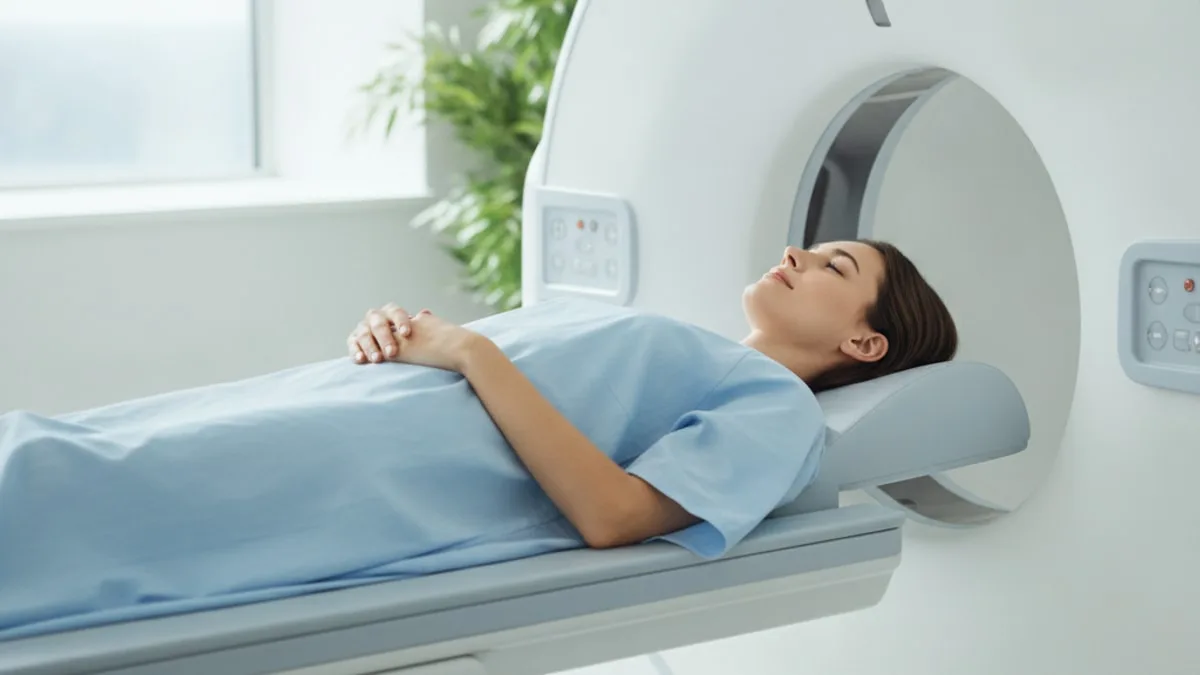
PET Scan: How It Works and What It's For
Everything below answers one question at a time, how the sugar signal works, what the test is used for, why fasting and blood sugar are part of the preparation, what the radiation exposure means in plain terms, and when a finding belongs in front of a clinician. This page is educational and is not medical advice.
What is a PET scan, and what does it measure?
A PET scan (positron emission tomography) is an imaging test that maps where a sugar-based scan preparation collects in the body, which reflects how actively tissue is using sugar rather than how that tissue is shaped.
That is the core difference from a plain X-ray, an ultrasound or a standard CT, which describe structure, size and density. Here the picture is built from a signal the body itself emits after the preparation is given. The preparation carries a small amount of radioactivity that decays and releases particles called positrons. Each positron meets an electron almost immediately, the two cancel out, and a pair of signals leaves the spot in opposite directions. A ring of detectors around the body registers those pairs, and software reconstructs a three dimensional map of where the signal came from and how concentrated it was.
The practical consequence is worth sitting with. A PET image can flag a change in how tissue behaves before, or alongside, a change in shape or size. It can also look quiet where tissue is structurally abnormal but metabolically slow. Reading one is a comparison exercise, the intensity in one place against the tissue around it, against the rest of the body, and against any earlier scan of the same person.
One thing the scan does not do is name a condition. It produces a map of signal intensity. What that map reflects is a judgement made by a radiologist or nuclear medicine physician who reads it together with your history, your examination and your other tests.
How does a sugar-based scan preparation make active tissue show up?
Cells that take up more sugar collect more of the preparation, and the molecule stays inside them long enough for the scanner to register it.
The preparation is a sugar-like molecule that enters cells through the same transporters glucose uses. Once inside, the first step of normal sugar processing happens to it, and then the altered molecule cannot continue down the usual pathway, so it builds up instead of being burned, a behaviour described as metabolic trapping 1. Research suggests that the intensity seen on the image reflects how quickly cells take up and hold the preparation, which is why the scan is described as a functional test rather than a structural one 1.
Several kinds of tissue use a lot of sugar. Brain tissue does. Heart muscle does under certain conditions. Activated white cells gathering at a site of infection or inflammation do. Cancer cells can also take up more sugar than the tissue around them 1. That overlap is the single most important thing a reader can understand about this test, because the scanner reports concentration, not cause. A bright area says that something there is working hard with sugar. It does not say why.
The body also clears the preparation, partly through the kidneys and bladder, so those structures can appear bright on an image for ordinary reasons rather than clinical ones 1. Experienced readers expect that pattern and account for it.
Why is the scan combined with a CT?
Because an activity map without an anatomical map is hard to place, the two are acquired together on a combined scanner during the same appointment.
The PET portion answers the question of where the signal is intense. The CT portion answers the question of what structure sits at that exact spot, a lymph node, a bone, a lung nodule, a loop of bowel. Combining them also serves a technical purpose, because the CT data are used to correct the PET signal for the way body tissues absorb it on its way to the detectors, which makes the activity map more faithful 1.
| Imaging test | What it mainly captures | What it contributes to a combined scan |
|---|---|---|
| PET | Where a sugar-based scan preparation collects, which reflects tissue activity | Flags areas of raised activity that structural images can miss |
| CT | Density and anatomy, including bone, lung and organ outlines | Pins the activity to a precise location and supports signal correction |
| MRI | Soft tissue detail and contrast between similar tissues | Used in combined systems where soft tissue definition is the priority |
You may see the result written as PET/CT on a report or a request form. Combined PET and MRI systems also exist and have been compared with PET/CT in some clinical questions, as described further below 9.
What is this imaging test used for?
Procedure guidelines for tumour imaging describe its use in cancer care for staging, for assessing response during or after therapy, for restaging and for investigating suspected recurrence 2.
Those four jobs cover much of the clinical demand. Staging asks how far a known cancer extends, including places a structural scan may not have reached. Response assessment asks whether activity in a known site has changed. Restaging asks what the current picture looks like after an interval. Recurrence asks whether a site that was managed earlier is active again, which can be difficult when scar tissue and changed anatomy make structural images ambiguous.
Beyond cancer, the same sugar signal is applied to questions about infection and inflammation, which the next sections cover with the specific settings that have been examined 789. Some heart and brain assessments also use positron emission imaging with other kinds of scan preparations, and the requesting clinician decides which approach fits the question being asked.
What this test is not is a general screening tool for people without symptoms or a reason to look. Decisions about screening, and about which imaging test suits a particular question, belong in a conversation with your clinician, who weighs the reason for looking against what the scan can and cannot answer.
How well does it pick up cancer that has come back?
It is used when there is a question of residual or recurrent disease after earlier management, and findings across several cancer types support that role, although imaging does not replace tissue sampling.
Research suggests that PET or PET/CT is useful for detecting residual or recurrent head and neck squamous cell carcinoma after earlier management 3. Studies suggest that it also contributes diagnostic information when recurrent cervical cancer is suspected 4. In renal cell carcinoma, research suggests that PET or PET/CT is informative when the clinical question is restaging 5. In pleural mesothelioma, research suggests that PET/CT carries diagnostic performance and clinical value in assessment 6.
Two caveats travel with all of that. The first is the overlap already described, because inflammation, healing tissue and infection can raise the signal in a way that looks similar to active tumour on the image alone 1. The second is the resolution limit, since a very small deposit of disease can sit below what the scanner can resolve, so a quiet image is not the same as an empty body.
That is why a bright area is treated as information rather than an answer. The clinician looking after you weighs it against your symptoms, your earlier imaging and your tumour markers, and decides whether a biopsy, a different scan or an interval repeat is the right next step. A value or finding outside what the report describes as expected is worth discussing with that clinician, who decides what it reflects.
Can it be used for infection and inflammation?
Yes, because the white cells that gather at an inflamed or infected site are metabolically busy and take up the sugar-based preparation 1.
Several specific settings have been examined. Research suggests that PET or PET/CT contributes to the assessment of suspected prosthetic joint infection, where distinguishing infection from mechanical loosening is the clinical difficulty 7. In suspected pyogenic native vertebral osteomyelitis, studies suggest that PET/CT sits alongside MRI and nuclear scintigraphy among the imaging options used to assess the spine 8. In inflammatory bowel disease, research suggests that both PET or PET/CT and PET/MR provide diagnostic information, with the comparison between the two examined directly 9.
The practical meaning for a reader is that an inflammatory cause and a malignant cause can produce similar brightness, so the report describes the pattern, the location and the intensity, and the clinician interprets it against the clinical story. Recent surgery, a healing wound, a recent infection or a site of ongoing irritation can all contribute to signal, which is one reason the imaging team asks about timing before the appointment 2.
If you are asked about infections, operations or procedures in the preceding weeks, that is not paperwork for its own sake. It changes how the reader interprets what they see.
Why does blood sugar matter before the appointment?
Because the preparation competes with the sugar already circulating in your blood for entry into cells, so a raised blood sugar can dampen the signal the scan depends on 1.
Think of the transporters on a cell surface as a limited number of doors. If a large amount of ordinary glucose is queueing at those doors, less of the scan preparation gets through, and the contrast between busy tissue and quiet tissue flattens. Raised circulating insulin after a meal adds another wrinkle, because it drives sugar and the preparation into muscle and fat, pulling signal away from the area of interest 1.
That is the whole reason for the fasting instruction. Procedure guidelines for tumour imaging describe fasting before the preparation is given and measuring blood glucose beforehand as part of standard preparation, with the scan reconsidered when the measured value sits too high for a reliable picture 2.
In people with diabetes, the imaging team plans the timing of the appointment around meals and medicines, and will ask what you take and when. Do not change anything yourself. Some medicines affect blood sugar, so ask your prescriber well before the appointment rather than on the morning of it, and follow the written instructions the imaging centre gives you.
Where does the fasting blood sugar number fit in?
The number the imaging team checks before your scan is the same quantity measured on a routine blood panel. Fasting Glucose is the blood sugar level measured after a period without food, and the parent guide covers what it measures, how it is tested and how to read a result.
FitScript's optimal range for Fasting Glucose is 75 to 86 mg/dL, an optimization target rather than a diagnostic cut-off, and your lab's own range appears as printed on your lab report. A value outside the printed range is worth discussing with a clinician, who decides what it reflects and whether anything needs following up. A single reading taken in a waiting room, after a stressful morning and a broken night, is a snapshot rather than a verdict.
How long does the whole appointment take?
Plan for the visit to run considerably longer than the scanning itself, because the preparation needs a quiet waiting period inside the body before images are acquired 2.
People asking how long a PET scan takes are thinking of the time on the table, which is the shorter part. The visit has a sequence. You check in and answer questions about fasting, medicines, pregnancy and recent illness or surgery. Your blood sugar is measured 2. The preparation is given by the imaging team. Then comes a quiet rest period in a warm room, where you are asked not to talk, chew, read aloud or move much, because muscle activity draws the preparation into working muscle and competes with the signal the scan is after 1. You will be asked to empty your bladder before the scan starts, since the preparation is cleared that way 1. Then the images are acquired while you lie still, with arm position and breathing instructions depending on the body area being examined.
Total time varies with the region being imaged, the equipment, whether extra images are needed afterwards and how busy the department is, so ask the centre for the expected duration when you book and arrange your day around that rather than around an internet estimate.
How should you prepare, and what should the imaging team know?
Follow the written instructions from the imaging centre exactly, because they exist to stop blood sugar and muscle activity from distorting the picture 2.
The usual elements of preparation look like this.
- A period without food before the appointment, as specified by the centre, with water as instructed 2.
- Avoiding hard exercise in the day or so beforehand, since working muscle takes up the preparation and can crowd the image 1.
- Staying warm on the day, because cold exposure activates brown fat, which is metabolically busy and can light up on the image 1.
- Resting quietly during the waiting period rather than talking, chewing or walking around 1.
- Emptying the bladder before images are acquired 1.
Tell the team in advance about pregnancy or the possibility of pregnancy, breastfeeding, diabetes, recent surgery, a recent infection, recent cancer therapy, difficulty lying flat, difficulty with enclosed spaces, and any mobility or weight considerations that affect positioning. Each of those changes either the scheduling, the interpretation or the safety discussion. For medicines, the instruction is simple. Some medicines matter here, so ask your prescriber what to do about yours, and do not stop or alter anything on your own.
How much radiation is involved, in plain terms?
The scan involves a small amount of ionising radiation from two sources, the sugar-based scan preparation given beforehand and the CT portion acquired with it.
The precise amount depends on the equipment, the body area covered, how the CT is configured and your size, so there is no single figure that fits every scan. Your imaging department can tell you the expected exposure for the scan you are booked for, and that is the right place to ask, rather than a general article.
What is useful to understand is where the exposure goes. The radioactivity in the preparation decays over a matter of hours, and what is left is cleared from the body, partly in urine, which is why staff ask you to drink fluids and empty your bladder afterwards 1. Departments give their own advice about close contact with infants, young children and pregnant people for the rest of the day, and that advice is specific to their protocol, so follow theirs.
In pregnancy or while breastfeeding, the conversation happens before scheduling, not on the day. The balance between the information the scan provides and the exposure it involves is a judgement the requesting clinician and the imaging team make together, with you, for your particular question.
What can make the images harder to read, and what is an incidental hot spot?
Plenty of ordinary things raise or lower the signal, which is why a report describes findings in context instead of naming a condition.
Normal tissue accounts for much of it. The brain, the heart under some conditions, the kidneys and bladder along the clearance route, and sections of bowel can all show activity for physiological reasons 1. Muscle that has been working takes up the preparation, as does brown fat in someone who got cold on the way in 1. Healing tissue after surgery, an active infection and inflammation of any cause can look bright 1. Pulling in the other direction, a raised blood sugar flattens contrast across the whole image 1, and a deposit smaller than the scanner can resolve may not register at all. Movement during acquisition blurs the map.
An incidental hot spot is activity found somewhere nobody was looking, which happens because the scan covers a wide field. These are assessed rather than dismissed. Research suggests that incidental uptake in the oropharynx detected on PET-CT carries a measurable risk of malignancy, which is why such a finding prompts further assessment rather than reassurance 10.
What an incidental finding is not is a diagnosis. It is an observation. The next step, which might be an examination, a different test, a sample or an interval repeat, is decided by the clinician looking after you.
What does the report tell you, and who decides what it means?
The report describes where the preparation collected, how intense that collection was, how it compares with surrounding tissue and with any earlier scan, and what the reader considers the finding is consistent with.
Reports can include a number expressing how concentrated the signal is relative to the rest of the body, used mainly to compare the same site across time rather than as a threshold that separates one condition from another 1. A conclusion written as consistent with, suspicious for or indeterminate is doing careful work. Those phrases mean the reader is describing a probability given the pattern, not naming a condition.
A useful way to read your own report is in three steps. First, the observation, which is what the scan measured and where. Second, the possible meanings, which is what the finding may reflect given everything else known about you. Third, the next step, which is what the clinician who ordered the scan recommends doing about it. Only the first of those is in the report itself.
Discuss your results and any proposed treatment with the clinician or licensed provider looking after you, and ask them directly what the finding changes about your plan. Treatment decisions belong to a licensed provider who has reviewed your full picture.
Which other tests can sit alongside imaging?
Imaging answers a structural and functional question at one moment, and blood work fills in the metabolic background that shapes both your preparation for the scan and your health more broadly.
Because the test runs on sugar handling, the blood markers describing sugar handling are the natural companions. Fasting glucose is the snapshot. HbA1c describes longer term blood sugar exposure. Fasting insulin describes how hard the body is working to hold glucose where it is. Alongside those, a clinician may look at kidney and liver panels, a complete blood count and inflammation markers such as CRP, which can be relevant when the question involves infection or inflammation. None of those replaces imaging, and imaging does not replace them.
A membership such as FitScript tests 150+ biomarkers per year, with interpretation by Atlas, its AI health advisor, and treatments decided on by a licensed provider after reviewing the member. The point of that pairing is context. A single glucose reading taken before a scan says much less than the same number read against the rest of your metabolic panel over time.
When is it worth talking to a clinician?
Any finding in a scan report, and any blood value outside the range printed on your lab report, is worth discussing with the clinician who ordered the test, because they decide what it reflects and what follows.
Call 911 or go to the emergency department for chest pain or pressure, trouble breathing, or fainting or passing out. Those signs are not for a waiting room call back or a later appointment.
Contact a clinician promptly, rather than waiting for a routine review, for a new or growing lump, unexplained weight loss, a persistent fever, drenching night sweats, bleeding from anywhere you cannot account for, or a symptom that has changed character since your last review. Bring the scan report itself to the conversation, along with your recent blood work, because the two read better together than apart.
If a therapy is considered after imaging, a licensed provider evaluates whether it is appropriate for you. Decisions about whether to scan at all, which test to use and how often to repeat it, are the same kind of conversation, shaped by your history rather than by a general rule.
Key takeaways
- A PET scan maps how actively tissue uses sugar, so it reports on function rather than shape alone, and it is acquired with a CT to show exactly where that activity sits 1.
- The signal depends on sugar handling, which is why fasting and a blood glucose check are part of standard preparation for tumour imaging 2.
- Procedure guidelines describe its use in staging, response assessment, restaging and suspected recurrence 2, and findings also support roles in prosthetic joint infection 7, vertebral osteomyelitis 8 and inflammatory bowel disease 9.
- Inflammation, healing tissue, working muscle, brown fat and the body's own clearance route can all brighten an image, so a hot spot is an observation rather than a diagnosis 1.
- Incidental uptake is assessed rather than ignored, since research suggests such findings can carry a risk of malignancy 10.
- Radiation exposure is small but not zero, and your imaging department can give the figure for your specific scan.
- Call 911 or go to the emergency department for chest pain or pressure, trouble breathing, or fainting.
References
- The Basic Principles of FDG-PET/CT Imaging. PET Clinics, 2014.
- FDG PET/CT: EANM procedure guidelines for tumour imaging: version 2.0. European Journal of Nuclear Medicine and Molecular Imaging, 2015.
- Detecting Residual/Recurrent Head Neck Squamous Cell Carcinomas Using PET or PET/CT: Systematic Review and Meta-analysis. Otolaryngology-Head and Neck Surgery, 2016.
- Diagnostic value of 18F-FDG-PET or PET-CT in recurrent cervical cancer: a systematic review and meta-analysis. Nuclear Medicine Communications, 2014.
- Diagnostic performance of 18F-FDG PET or PET/CT in restaging renal cell carcinoma: a systematic review and meta-analysis. Nuclear Medicine Communications, 2017.
- The diagnostic performance and clinical value of 18F-FDG PET/CT in pleural mesothelioma, a systematic review and meta-analysis. Seminars in Nuclear Medicine, 2026.
- Diagnostic role of PET or PET/CT for prosthetic joint infection: A systematic review and Meta-analysis. Hellenic Journal of Nuclear Medicine, 2021.
- The use of MRI, PET/CT, and nuclear scintigraphy in the imaging of pyogenic native vertebral osteomyelitis: a systematic review and meta-analysis. The Spine Journal, 2023.
- Comparing the Diagnostic Value of FDG PET or PET/CT With FDG PET/MR in Inflammatory Bowel Disease, A Systematic Review and Meta-analysis. Clinical Nuclear Medicine, 2024.
- Prevalence and malignant risk of incidental uptake in the oropharynx detected by PET-CT: a systematic review and meta-analysis. European Archives of Oto-Rhino-Laryngology, 2025.
Common questions
What is a PET scan used for?
It is used in cancer care to assess how far a known cancer extends, to look at how a site has responded after therapy, and to investigate whether disease has become active again. The same sugar signal is also applied to questions about infection and inflammation, such as a suspected infection around a joint replacement or in the spine. The clinician who orders it decides whether it is the right test for the question being asked.
How long does a PET scan take?
The time on the scanner is the shorter part of the visit. Most of the appointment is taken up by check-in questions, a blood sugar check, and a quiet rest period after the preparation is given, before any images are acquired. Duration varies with the body area, the equipment and the department, so ask the imaging centre for the expected total when you book.
Do you have to fast before a PET scan?
Fasting is part of standard preparation, and the imaging centre will give you written instructions covering how long and what you may drink. The reason is that food raises blood sugar, and circulating sugar competes with the scan preparation for entry into cells, which flattens the picture. Your blood sugar is measured before the preparation is given, and the team may reschedule if the value is too high for a reliable scan.
Does a PET scan hurt?
The imaging itself is painless, and you simply lie still on a table while the scanner moves around you. Having the preparation given involves a needle, which can sting briefly. Lying flat and still can be uncomfortable for some people, so tell the staff beforehand if you cannot lie flat or find enclosed spaces difficult.
How much radiation does a PET scan involve?
It involves a small amount of ionising radiation from two sources, the scan preparation and the CT portion acquired with it. The exact amount depends on the equipment, the body area covered and how the scan is set up, so your imaging department can tell you the figure for your appointment. The clinician requesting the scan weighs the information it provides against the exposure, and that conversation should happen before scheduling in pregnancy or while breastfeeding.
Can a PET scan tell you whether a spot is cancer?
No. It shows where tissue is taking up sugar intensely, and inflammation, infection, healing tissue and working muscle can all produce a similar bright area. A finding on the scan is an observation that sits alongside your symptoms, your examination and your other tests. The clinician looking after you decides whether a biopsy, a different test or a repeat scan is the next step.
What happens if the scan shows an unexpected hot spot?
An incidental hot spot is an area of activity found somewhere nobody was specifically looking, which happens because the scan covers a wide field. It is assessed rather than set aside, and the next step may be an examination, another imaging test, a tissue sample or a repeat scan after an interval. Who decides is straightforward, it is the clinician who ordered the scan, working from the full report.
Can people with diabetes have a PET scan?
Yes, and the imaging team plans the appointment around meals and medicines because blood sugar affects the quality of the picture. Your blood sugar is measured before the preparation is given, and the team may adjust the timing or reschedule based on what they find. Do not change or stop any medicine on your own, ask your prescriber well before the appointment and tell the imaging centre what you take.