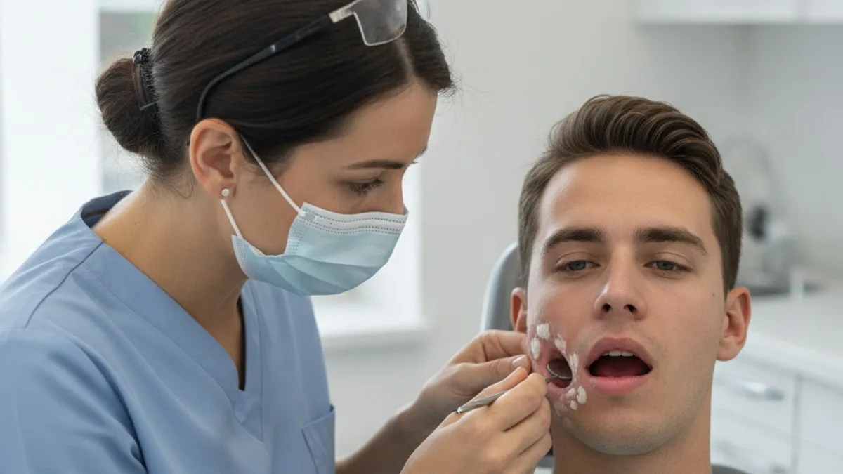
Oral Leukoplakia Symptoms: White Patches in the Mouth
A mouth sore with heavy bleeding, or swelling in the mouth, tongue or throat that makes it hard to breathe or swallow, means call 911 or go to the emergency department.
Everyday things change the mouth lining as well. Friction from a sharp tooth or a denture, cheek chewing, tobacco, alcohol, vaping, dry mouth and some medicines all come up in that conversation, and your prescriber is the person to ask about any medicine you take. Blood work plays a supporting role here. A complete blood count, ferritin, vitamin B12, folate, HbA1c and vitamin D describe the background the mouth lining is working in, and a clinician decides what any result reflects.
What are the symptoms of oral leukoplakia?
The main symptom of oral leukoplakia is a white or greyish white patch on the lining of the mouth that stays in place when you rub, wipe or brush it. Clinical descriptions of oral potentially malignant disorders note that these patches appear on the tongue, the inner cheek, the gums, the lip and the floor of the mouth 1. Some are flat, thin and even in colour. Others are thickened, wrinkled or raised, or carry red areas mixed into the white, an appearance clinicians describe as non-homogeneous 1.
The patch can carry no sensation at all, which is part of why it gets noticed late or by a dentist rather than by the person. When something is felt, people describe a rough area the tongue keeps returning to, a leathery or firm patch, tenderness, burning with spicy or acidic food, or a spot that catches during chewing. A patch can also change over months, becoming thicker or developing a different texture across part of its surface.
Oral leukoplakia is the name used once other explanations for a white patch have been ruled out, so it is a description reached by exclusion rather than by looking alone 2. Research suggests oral potentially malignant disorders, the group that includes leukoplakia, are found in adult populations around the world 3. Studies suggest that a share of oral leukoplakia lesions change into oral cancer over time, which is the reason a patch that persists is examined rather than watched from home 4.
What you can observe is limited to the surface. Note where the patch sits, how long it has been there, whether it has grown or changed colour, whether the surface has turned rough or hard, and whether it bleeds when touched. Those details can reflect friction, an irritant the lining is reacting to, or a change in the cells themselves, and the surface alone does not separate them. The next step is an examination by a dentist or an oral medicine specialist, who decides whether a biopsy is needed.
What can cause white patches in the mouth?
Several everyday exposures can change the colour and texture of the mouth lining, and not all white patches are leukoplakia. Friction is one of them. Repeated rubbing from a sharp tooth edge, a broken filling, a denture that no longer fits well, or habitual cheek and lip chewing can thicken an area of lining over weeks and months.
Habits and intake matter too. Tobacco in smoked and smokeless forms, areca nut and betel quid, and alcohol are described in clinical reviews as risk factors for oral potentially malignant disorders 2. Research suggests e-cigarette use may be a risk factor for oral potentially malignant disorders and oral cancer 5. A dentist asks about all of these because the exposure history shapes how closely a patch is followed.
Other day-to-day factors change how the lining looks and feels without being leukoplakia at all. A white film that comes away when wiped behaves differently from a patch that stays. A dry mouth from mouth breathing at night, from heavy training without enough fluid, or as a side effect of some medicines leaves the lining more easily irritated, and your prescriber is the person to ask about any medicine you take. Stress-driven clenching and cheek chewing can leave a ridged pale line along the level where the teeth meet. Poor sleep, a heavy training block and a diet low in fresh food do not create a white patch on their own, but they sit in the background of how quickly the lining renews itself.
None of these causes show up on a blood panel. They are found by asking questions and by looking in the mouth, which is why the examination leads and the bloods follow.
How is a patch that stays assessed, and why does a biopsy matter?
A clinician examines the whole mouth, records where the patch is and how long it has been present, removes sources of friction where that is possible, and takes a biopsy when a patch remains. Consensus guidelines on the management of oral potentially malignant disorders describe biopsy as the step that establishes what the tissue actually is 6. A biopsy takes a small sample so a pathologist can look at the cells and report whether dysplasia is present and to what degree 2.
That report is the piece no visual check can supply. Studies suggest that lesions with a non-homogeneous appearance and with higher grades of dysplasia carry a greater chance of change into oral cancer 4. A review from the American Head and Neck Society Cancer Prevention Service sets out current clinical management of oral leukoplakia, including removal of the lesion where appropriate and continued surveillance afterwards 7.
Follow-up is part of the picture rather than an optional extra. Guidance on management describes continued review of people with oral potentially malignant disorders over time 6, and the management update describes surveillance after a lesion has been removed because patches can return or appear elsewhere 7. In practice that means a named dentist or specialist who sees the same mouth at intervals and can tell what has changed. Treatment decisions belong to a licensed provider.
What is proliferative verrucous leukoplakia?
Proliferative verrucous leukoplakia, written PVL, is a form in which white patches appear at more than one site in the mouth and spread over time, with surfaces that become wrinkled or wart-like 8. An expert consensus guideline sets out how PVL should be assessed and reported so that clinicians describe the same thing in the same terms 8.
The distinction matters because PVL is associated with a higher chance of change into cancer than a single localised patch 8. It is also harder to judge from one appointment, since the defining feature is behaviour over time rather than a single appearance.
Which blood tests can help explain mouth symptoms?
No blood test identifies a white patch in the mouth, so blood work sits beside the oral examination rather than replacing it. Where blood work earns its place is in the background. The lining of the mouth renews quickly, and markers of iron status, B vitamins, blood sugar and general blood counts describe the conditions that renewal happens in. A smooth or burning tongue, cracks at the corners of the mouth and ulcers that keep returning are the kinds of complaints that prompt a clinician to look at these markers.
| Marker | Why a clinician can consider it | What a result can and cannot tell you |
|---|---|---|
| Complete blood count (CBC) | Describes red cells, white cells and platelets, and gives a first view of oxygen carrying and immune activity | Can show a pattern worth following up for iron or B vitamin status. Cannot describe the tissue in a white patch |
| Ferritin | Reflects stored iron, which the fast-renewing mouth lining draws on | Can flag low stores behind a smooth or tender tongue. Rises with inflammation, so one value does not settle the question |
| Vitamin B12 | Needed for cell division in tissues that renew quickly | Can explain a sore, red or burning tongue and recurring ulcers. Says nothing about a patch that will not wipe off |
| Folate | Works alongside B12 in cell division and moves with diet and alcohol intake | Can add context to mouth soreness. Cannot grade or exclude dysplasia |
| HbA1c | Reflects average blood glucose over the previous few months | Can show whether blood sugar is part of the background to a dry mouth or surface overgrowth. Does not describe a lesion |
| Vitamin D (25-hydroxyvitamin D) | Takes part in immune function and tissue maintenance | Can show whether status is low. A low value is not an explanation for a white patch |
Ranges are not printed here on purpose. Read each value against the range as printed on your lab report, and a value outside that range is worth discussing with a clinician, who decides what it reflects.
Can a complete blood count explain mouth changes?
A complete blood count can explain some mouth complaints, but never a white patch. It measures red cells, white cells and platelets, so it shows whether the blood is carrying oxygen well and whether the immune side of the count is doing something unusual. Low haemoglobin alongside small red cells points a clinician toward iron status, and that pattern can sit behind tiredness, breathlessness on stairs and a tongue that looks smoother than it used to.
What a CBC cannot do is see the lining. It reports cells circulating in the blood, not the cells stacked in a patch on the cheek or tongue. If a value sits outside the range printed on your lab report, that is worth discussing with a clinician, who decides what it reflects and whether further testing is sensible. It does not change the need for a dentist to look at a patch that stays.
Can low ferritin affect the tongue and mouth?
Low ferritin can be one reason for a sore, smooth or tender tongue, though it is not a reason for a patch that will not wipe off. Ferritin reflects stored iron. Cracks at the corners of the mouth, a tongue that has lost some of its texture and general fatigue are the kinds of complaints that bring ferritin into the conversation.
Several things move ferritin at once. Heavy menstrual bleeding, low dietary intake, endurance training and problems with absorption in the gut all pull it down, while inflammation pushes it up, so a value inside the printed range alongside active inflammation does not close the question. A clinician can add other iron markers to read it properly. A ferritin result outside the printed range is worth discussing with a clinician rather than acted on alone.
What do vitamin B12 and folate have to do with a sore mouth?
Vitamin B12 and folate are needed for cell division, and the mouth lining divides quickly. People describe a red, smooth and sore tongue, a burning feeling across the lining, and ulcers that keep coming back in the same places.
Low B12 can follow low intake, reduced absorption with age, gut surgery, conditions affecting the stomach lining, and some medicines, which is a question for your prescriber rather than for a search engine. Folate moves with diet and with alcohol intake. Neither marker describes a white patch, and a normal result for both does not make a persistent patch less worth examining. Values outside the ranges printed on your lab report are worth discussing with a clinician, who decides what they reflect.
Does blood sugar come into this?
Blood sugar is part of the background to mouth symptoms rather than an explanation for leukoplakia. HbA1c reflects average blood glucose over the previous few months, while a fasting glucose captures a single moment. Higher glucose can go with a drier mouth and with overgrowth on the lining, and a surface overgrowth can look white while behaving differently from a patch that stays fixed in place.
In people with higher blood sugar, dentists pay close attention to the gums and the lining at routine visits, since healing and gum health shift with glucose control. If HbA1c or fasting glucose sits outside the range printed on your lab report, that is worth discussing with a clinician promptly, who decides what it reflects and what comes next.
Where does vitamin D fit in?
Vitamin D fits in as background rather than as a cause of a white patch. Labs use 25-hydroxyvitamin D as the marker of status, and vitamin D takes part in immune function and in the maintenance of bone and other tissues. A low value does not explain a patch, and bringing it back into range does not remove the need for the patch to be examined.
It still earns a place on a panel because it is easy to be low without noticing and because it sits alongside the other markers that describe how well the body is supporting fast-renewing tissue. A value outside the printed range is worth raising with a clinician, who decides whether anything needs to change.
What can a normal panel still miss?
A full set of in-range results says nothing at all about what the tissue under a white patch looks like.
Blood work also misses the mechanical and habit side of the story. Patches that appear at several sites and spread need the structured assessment set out for PVL rather than a single snapshot 8.
The honest summary is that bloods describe the body around the mouth. The mouth itself is checked by eye, by hand and, when a patch stays, by biopsy.
When should you see a clinician quickly?
Some mouth signs need emergency care and some need a prompt appointment, and they are not the same list.
Call 911 or go to the emergency department for any of these.
- Heavy bleeding from a mouth sore or from the gums that does not stop with pressure
- Swelling in the mouth, tongue or throat that makes it hard to breathe or swallow
- Trouble breathing
- Chest pain or pressure
- Fainting or passing out
Arrange a visit with a dentist or doctor without waiting for the next routine check-up for any of these.
- A white patch that will not wipe off and has been present for more than two weeks
- A patch that has grown, thickened, hardened or developed red areas within it
- A mouth ulcer or sore that has not healed
- A patch that bleeds when touched lightly
- A new lump in the mouth, the lip or the neck
- Numbness or constant tenderness in one area of the mouth
- A tooth that has loosened without an injury
- A patch on the floor of the mouth, under the tongue or along the side of the tongue
Do not try to settle what a patch is from a photo or a search result. A patch that stays is a reason for an examination, and the clinician decides whether a biopsy is needed and what any finding means.
If you are having thoughts of self-harm, call or text 988 (US) or your local emergency number now.
How can you check these markers?
The FitScript Essential panel measures 40+ biomarkers.
Atlas, FitScript's AI health advisor, reads those results back in plain language, explains what each marker describes and flags what is worth raising at an appointment.
Blood work does not replace an oral examination. A white patch is assessed by a dentist or an oral medicine specialist, a biopsy describes the tissue, and treatment decisions belong to a licensed provider after reviewing the person in front of them. Discuss your results and any treatment with a clinician or licensed provider before changing anything. This page is educational and not medical advice.
Key takeaways
- The core sign of oral leukoplakia is a white or greyish patch on the lining of the mouth that stays put when rubbed or brushed, and it can be painless.
- Clinical descriptions note that flat, even patches and thickened or mixed red and white patches look different and are followed differently 1.
- No blood test identifies a white patch. Guidelines describe biopsy as the step that establishes what the tissue is 6.
- A complete blood count, ferritin, vitamin B12, folate, HbA1c and vitamin D describe the background and can explain a sore or smooth tongue, not a patch.
- Heavy bleeding from a mouth sore, or swelling that makes it hard to breathe or swallow, means call 911 or go to the emergency department.
References
- Clinical features and presentation of oral potentially malignant disorders. Oral Surgery, Oral Medicine, Oral Pathology and Oral Radiology, 2018.
- Oral potentially malignant disorders: A comprehensive review on clinical aspects and management. Oral Oncology, 2020.
- Prevalence of oral potentially malignant disorders: A systematic review and meta-analysis. Journal of Oral Pathology & Medicine, 2018.
- Malignant transformation of oral leukoplakia: Systematic review and comprehensive meta-analysis. Oral Diseases, 2025.
- The use of E-cigarettes as a risk factor for oral potentially malignant disorders and oral cancer: a rapid review of clinical evidence. Medicina Oral, Patologia Oral y Cirugia Bucal, 2024.
- Consensus guidelines on management of oral potentially malignant disorders. Indian Journal of Cancer, 2022.
- Clinical Management Update of Oral Leukoplakia: A Review From the American Head and Neck Society Cancer Prevention Service. Head & Neck, 2025.
- Proliferative Verrucous Leukoplakia: An Expert Consensus Guideline for Standardized Assessment and Reporting. Head and Neck Pathology, 2021.
Common questions
What are the causes of leukoplakia?
Leukoplakia is the name given to a white patch once other explanations have been excluded, and clinicians look at exposures that irritate the lining. Tobacco in smoked and smokeless forms, areca nut and betel quid, alcohol, e-cigarette use and friction from a sharp tooth, a broken filling, a poorly fitting denture or cheek chewing all come into that assessment.
Can a white patch in the mouth be wiped off?
A leukoplakia patch stays in place when you rub, wipe or brush it. A white film that comes away with gauze and leaves the surface underneath behaves differently and points a clinician in another direction. Either way, a dentist is the person who examines the patch and decides what happens next.
Does oral leukoplakia hurt?
A patch can sit in the mouth with no sensation at all, which is part of why it gets noticed at a dental check rather than by the person. When something is felt, people describe roughness, a leathery or firm area, tenderness, or burning with spicy and acidic food. Pain is not what decides whether it needs assessing.
How long should a white patch last before I see a dentist?
Arrange a dental visit if a white patch that will not wipe off has been there for more than two weeks, and sooner if it has grown, thickened, hardened, developed red areas or started to bleed when touched. Removing an obvious source of rubbing is part of the assessment, not a reason to wait and watch at home.
Can a blood test identify leukoplakia?
No. Blood tests describe what is circulating in the blood, not the cells in the lining of the mouth. A white patch is assessed by examination and, when it persists, by a biopsy that a pathologist reads. Blood work runs alongside that to describe general background health.
Which blood markers might a clinician check alongside an oral examination?
A complete blood count, ferritin, vitamin B12, folate, HbA1c and vitamin D are the markers that come up when someone has a sore, smooth or burning tongue, cracks at the corners of the mouth or ulcers that keep returning. Read each result against the range printed on your lab report and discuss anything outside it with a clinician.
Does vaping matter when a white patch is assessed?
Clinicians ask about vaping as well as smoking and smokeless tobacco when they assess a white patch, because the lining of the mouth is exposed to what passes over it. Tell the dentist what you use and how much, since the exposure history shapes how closely a patch is followed and whether a biopsy is taken.
What happens during a biopsy of a mouth patch?
A clinician numbs the area and removes a small sample of the patch, which a pathologist examines under a microscope and reports on, including whether dysplasia is present and to what degree. That report guides what the dentist or specialist does next. Any decision about removal or further care belongs to a licensed provider.