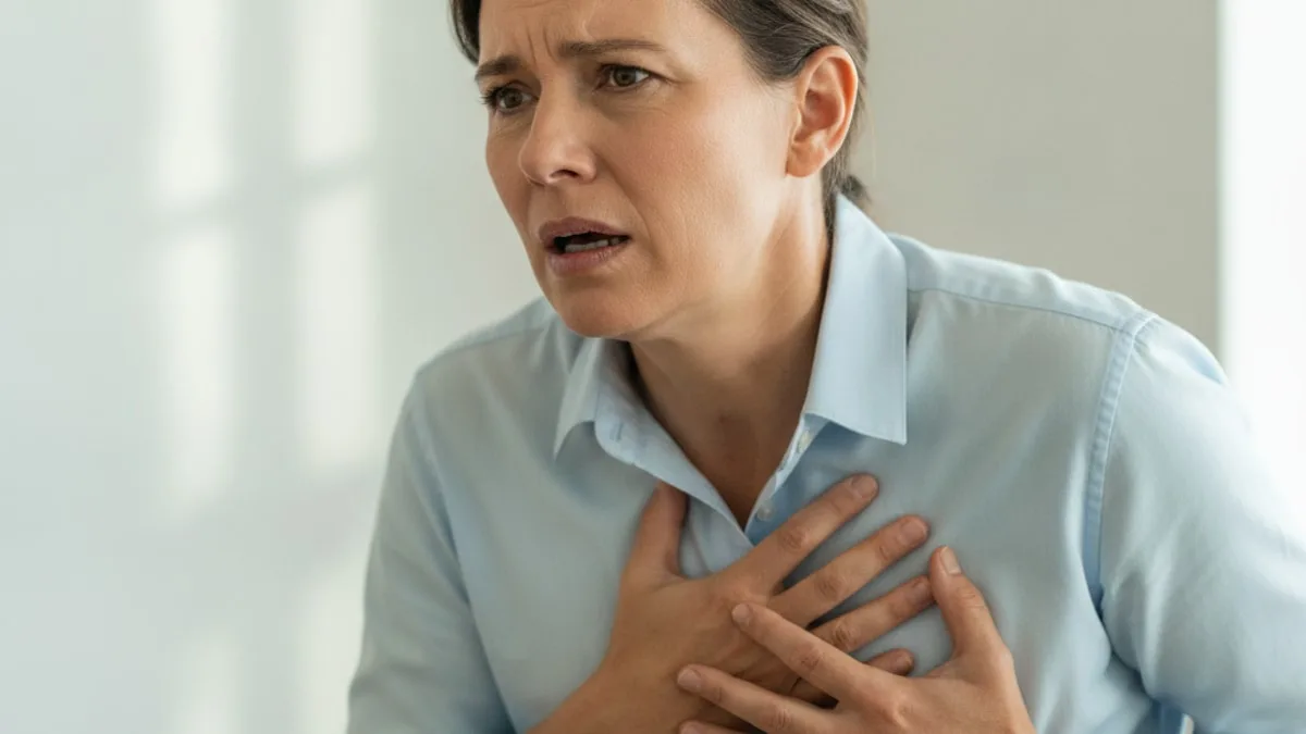
Pulmonary Edema: Causes of Fluid in the Lungs
Severe breathlessness, breathlessness that starts or gets worse when lying flat, or a cough that brings up pink frothy fluid needs emergency care. Call 911 or go to the emergency department.
The rest of this page explains where the fluid comes from, what each blood marker can and cannot tell you, what clinicians look for on a chest X-ray, and the signs that need emergency care.
What causes fluid to collect in the lungs?
Fluid collects in the lungs when pressure inside the lung blood vessels rises high enough to push water into the air spaces, or when the thin barrier between those vessels and the air sacs starts to leak.
Breathlessness on its own has many everyday explanations. Short sleep, a stressful stretch at work, a sudden jump in training load, alcohol the night before, a chest infection, extra body weight and low iron stores all make breathing feel like harder work. None of those is fluid in the lungs, and none of them is settled by a single blood value.
When fluid really has gathered in the lungs, the causes fall into two groups. Research suggests that pulmonary edema develops either from raised pressure in the lung capillaries or from a more permeable barrier between the capillary and the air sac, and the two patterns can overlap in the same person 1.
Heart-related causes sit in the pressure group. When the left side of the heart cannot move blood forward well, pressure backs up through the lung veins and water is pushed out into lung tissue. Clinical practice guidance on acute pulmonary edema describes this rapid rise in left-sided heart pressure as the route to breathlessness that comes on over minutes to hours 2. Worsening valve function, a heart rhythm that will not settle, uncontrolled blood pressure and a heart muscle that has been under strain for years all push in the same direction.
Non-heart causes work through leak rather than pressure. They include the following.
- Rapid ascent to high altitude. Research suggests that an uneven and exaggerated rise in lung artery pressure under low oxygen is associated with high altitude pulmonary edema 3. A scoping review examined high altitude pulmonary edema in women and set out recommendations for women travelling to altitude 4. A systematic review also described high altitude pulmonary edema in children who travel to altitude 5. Ask your child's pediatrician before taking a child to high elevation.
- Smoke inhalation. Evidence suggests that pulmonary edema after toxic smoke inhalation can begin hours after the exposure rather than straight away, so a person who feels reasonable at first can deteriorate later 6.
- Severe lung injury and critical illness. A narrative review of pulmonary edema in acute respiratory distress syndrome describes lung water as a leak-driven problem that is measured and followed closely in intensive care 7.
- Kidney problems. Kidneys set how much salt and water leave the body, so reduced filtration shifts the whole fluid balance.
- Severe infection, near drowning and chest injury, all of which can damage the barrier between blood vessel and air sac.
- Some medicines can change fluid balance or irritate lung tissue. Ask your prescriber about anything you take, and do not stop anything on your own.
- Pregnancy raises blood volume and heart workload. Ask your OB or midwife about new breathlessness in pregnancy.
More than one cause can be present at once. A heart under strain plus a heavy salt and fluid load plus a chest infection is a very different picture from any one of those alone, which is why a clinician looks at the history, the examination, imaging and blood work together rather than at a single result.
Which blood tests can help explain it?
No blood test shows fluid sitting in the lungs, but a short panel can show how the kidneys are filtering, how water and salt are balanced, and how well the blood is carrying oxygen.
| Marker | Why it is linked to this symptom | What a result can and cannot tell you |
|---|---|---|
| Sodium | Sodium concentration reflects the balance between body water and body salt, which shifts when fluid is being retained or lost. | Can flag that water and salt handling has drifted, which is worth discussing with a clinician. Cannot show whether the lungs hold fluid, and cannot name a cause on its own. |
| Creatinine | Creatinine reflects kidney filtration, and the kidneys decide how much salt and water the body can clear. | Can suggest that filtration has fallen, which affects fluid balance. Cannot separate a kidney cause from a heart cause, and shifts with muscle mass and hydration. |
| Hemoglobin | Hemoglobin carries oxygen, so a low value raises breathing effort and heart workload for the same task, while a high value can follow long-term low oxygen. | Can explain part of why breathing feels harder. Cannot show lung water, and changes with iron status, hydration, blood loss and altitude living. |
Read any value against the range printed on your own lab report, since reference ranges vary by lab, age and sex. A value outside that printed range is worth discussing with a clinician, who decides what it reflects.
In a hospital assessment of sudden breathlessness, clinicians add a natriuretic peptide test, written as BNP, which rises when the heart chambers are stretched by pressure or volume. It is interpreted in words rather than in isolation, alongside the examination and imaging, and it is one input among several rather than an answer by itself.
Can sodium levels explain swelling and breathlessness?
Sodium does not measure lung water, but it is one clue to how the body is holding on to fluid. The number on the report is a ratio rather than a total, because it describes how much sodium sits in a given volume of water.
That is why a low sodium value can appear when the body retains water faster than salt, a pattern seen in people with heart or kidney strain. Clinical practice guidance on acute pulmonary edema describes an assessment that combines history, examination, chest imaging and blood work rather than resting on any single number 2.
Plenty of ordinary things move this marker. Heavy fluid intake around long endurance sessions, hot weather, a stomach bug, a very low salt intake and some medicines all shift it, so ask your prescriber before changing anything you take. If sodium sits outside the range printed on your report, that is worth discussing with a clinician promptly rather than ignoring or self-correcting.
You can read more about what this marker measures on the Sodium page.
What can creatinine tell you about fluid balance?
Creatinine is a window on kidney filtration, and kidney filtration sets the ceiling on how much salt and water the body can remove each day. When that ceiling drops, fluid has fewer places to go.
A rising creatinine in someone with new breathlessness and swelling is one possible reason the balance has tipped, and it is a result a clinician will want to see alongside the rest of the picture.
Other things move creatinine without any kidney problem. More muscle mass pushes it up, dehydration pushes it up for a day or two, a large meat intake can nudge it, and a very hard resistance session the day before a draw can do the same. Age and some medicines also change it, so ask your prescriber about anything you are taking. A single raised value is worth repeating and discussing rather than acting on alone.
The Creatinine page explains what the marker measures and what moves it.
Can low hemoglobin make breathlessness worse?
Low hemoglobin means each unit of blood carries less oxygen, so breathing rate and heart rate climb for the same workload. That can make breathlessness noticeably worse, and it can sit on top of another cause rather than replacing it.
Oxygen supply matters in the other direction too. A hemoglobin that runs high can follow months of living at altitude or long-term low oxygen overnight, and that pattern is worth raising with a clinician rather than reading as a fitness marker.
Hemoglobin also moves for reasons that have nothing to do with the lungs. Iron intake and absorption, menstrual blood loss, slow gut bleeding, recent heavy endurance training that expands plasma volume, dehydration at the time of the draw and pregnancy all change it. Ask your OB or midwife about hemoglobin results in pregnancy.
See the Hemoglobin page for what the marker measures and the common reasons it drifts.
What can a normal panel still miss?
A great deal. Blood tests describe the chemistry around the lungs, not the water inside them, so a clean panel does not settle the question.
Imaging is where lung fluid becomes visible. On a chest X-ray, clinicians look for larger blood vessels in the upper parts of the lungs, thin horizontal lines near the lung edges known as Kerley B lines, cuffing around the airways, haze spreading outwards from the centre of the lungs and fluid collecting at the bases. A review of pulmonary edema pathophysiology and diagnosis describes chest radiography as a central part of recognising the pattern 1. Research suggests that lung ultrasound and other noninvasive imaging can quantify lung congestion more finely than a plain film describes 8.
Several other causes sit outside both the panel and the X-ray. Heart valve function and heart muscle performance need an ultrasound of the heart. Breathing that stops and starts during sleep needs a sleep test. Rhythm problems that come and go need monitoring over time rather than a single reading. Airway and lung disease needs breathing tests. Exposure history matters as well, since evidence suggests that pulmonary edema following toxic smoke inhalation can be delayed, so the time of exposure is part of the assessment 6.
Timing is the last gap. A panel drawn on a calm day describes that day. It does not describe what happens during a hard effort, during an illness, during a long flight or at 4,000 metres. If breathlessness keeps appearing in a specific situation, note when it happens and what you were doing, and bring that record to the appointment.
When should you see a clinician quickly?
Some breathing symptoms cannot wait for an appointment. Call 911 or go to the emergency department for any of the following.
- Severe or rapidly worsening breathlessness.
- A cough that brings up pink or frothy fluid.
- Breathlessness that starts or worsens when lying flat, or that wakes you gasping.
- Blue or grey lips, face or fingertips.
- Chest pain, chest pressure or a feeling of crushing tightness.
- Confusion, drowsiness or difficulty staying awake.
- Fast, laboured breathing that does not settle at rest.
- Breathlessness and cough that come on after going quickly to high altitude.
Other signs are not emergencies but should not be left to drift. Book a prompt appointment with a clinician for any of these.
- Breathlessness on effort that is clearly worse than it was a few weeks ago.
- New swelling in the ankles, feet or lower legs.
- Needing more pillows than usual to sleep comfortably.
- Unexplained weight gain over a handful of days.
- A cough that is worse at night.
- A lab value outside the range printed on your report, which is worth discussing promptly.
Do not try to work out the cause yourself from one number or one symptom. A clinician examines you, looks at imaging and reads your results together, and decides what they reflect.
If you are having thoughts of self-harm, call or text 988 (US) or your local emergency number now.
How can you check these markers?
Sodium, creatinine and hemoglobin are all measured in the FitScript Essential panel, so the three markers discussed above come back on one report rather than through three separate requests. Membership covers 150+ biomarkers tested per year, which means these values can be followed over time instead of read once in isolation.
Atlas, FitScript's AI health advisor, explains what each result means in plain language and shows how it sits against the range printed on the report. Interpretation is not the same as a decision. A licensed provider evaluates the whole picture and decides on any next step, including whether imaging or further assessment is appropriate.
This page is educational and is not medical advice. Discuss your results and any treatment with a clinician or licensed provider, since treatment decisions belong to a licensed provider.
Key takeaways
- Severe breathlessness, breathlessness lying flat and a pink frothy cough need emergency care. Call 911 or go to the emergency department.
- Research suggests that lung fluid comes either from raised pressure in the lung capillaries or from a leakier capillary barrier, and the two can overlap 1.
- Non-heart causes include rapid ascent to altitude, smoke inhalation and severe lung injury, and evidence suggests that the picture after toxic smoke inhalation can be delayed by hours 6.
- Sodium, creatinine and hemoglobin describe fluid balance, kidney filtration and oxygen carrying. None of them shows lung water directly.
- Chest imaging is where fluid becomes visible, and research suggests that lung ultrasound can quantify congestion beyond a plain film 8.
- A value outside the range printed on your report is worth discussing with a clinician, who decides what it reflects.
References
- Pulmonary edema: pathophysiology and diagnosis. The International Journal of Tuberculosis and Lung Disease, 2011.
- Clinical practice. Acute pulmonary edema. The New England Journal of Medicine, 2005.
- Pulmonary hemodynamics: implications for high altitude pulmonary edema (HAPE). A review. Advances in Experimental Medicine and Biology, 1999.
- High-Altitude Pulmonary Edema in Women: A Scoping Review-UIAA Medical Commission Recommendations. High Altitude Medicine & Biology, 2023.
- High altitude pulmonary edema in children: A systematic review. Pediatric Pulmonology, 2023.
- Delayed onset pulmonary edema following toxic smoke inhalation; a systematic review. Acute Medicine, 2018.
- Measuring and managing pulmonary edema in ARDS: a narrative review. Critical Care, 2026.
- Noninvasive Imaging Methods for Quantification of Pulmonary Edema and Congestion: A Systematic Review. JACC. Cardiovascular Imaging, 2023.
Common questions
pulmonary oedema
Pulmonary oedema is the same condition as pulmonary edema, written with the spelling used outside the United States. Both terms describe fluid collecting in the air spaces and tissue of the lungs, which makes breathing feel like hard work. Severe breathlessness, breathlessness lying flat or a pink frothy cough needs emergency care.
pulmonary edema clinical manifestations
The clinical manifestations include breathlessness that worsens lying flat, waking at night short of breath, fast and laboured breathing, a cough that can bring up pink frothy fluid, crackling sounds in the chest, anxiety, sweating and sometimes blue or grey lips. These signs need emergency care. Call 911 or go to the emergency department.
pulmonary edema on chest x ray
On a chest X-ray, clinicians look for larger blood vessels in the upper lungs, thin horizontal lines near the lung edges called Kerley B lines, cuffing around the airways, haze spreading outwards from the centre and fluid at the lung bases. A radiologist and clinician read the film together with your history.
What is the difference between heart-related and non-heart causes?
Heart-related fluid comes from pressure. When the left side of the heart cannot move blood forward, pressure backs up into the lung vessels and pushes water out. Non-heart causes come from leak, where the barrier between blood vessel and air sac becomes permeable, as with altitude exposure, smoke inhalation or severe lung injury.
Can a blood test on its own show fluid in the lungs?
No. Blood tests describe the chemistry around the lungs rather than the water inside them. They can show how the kidneys are filtering, how salt and water are balanced and how much oxygen the blood can carry. Imaging of the chest is what makes the fluid itself visible to a clinician.
Which blood markers might a clinician check?
Sodium, creatinine and hemoglobin are checked when breathlessness and swelling are being assessed. Sodium reflects water and salt balance, creatinine reflects kidney filtration, and hemoglobin reflects oxygen carrying capacity. A clinician may also order a natriuretic peptide test, which rises when the heart chambers are stretched.
Why does breathlessness get worse when lying flat?
Lying flat redistributes fluid from the legs and abdomen back towards the chest, which raises pressure in the lung circulation. People notice it as needing extra pillows or waking short of breath. Breathlessness that starts or worsens when lying flat needs emergency care. Call 911 or go to the emergency department.
Can going to high altitude cause fluid in the lungs?
Yes, a fast ascent to high elevation can lead to fluid in the lungs, which clinicians call high altitude pulmonary edema. Cough, severe breathlessness and poor exercise tolerance after going quickly to altitude need emergency care. Call 911 or go to the emergency department, and plan ascents with a clinician beforehand.