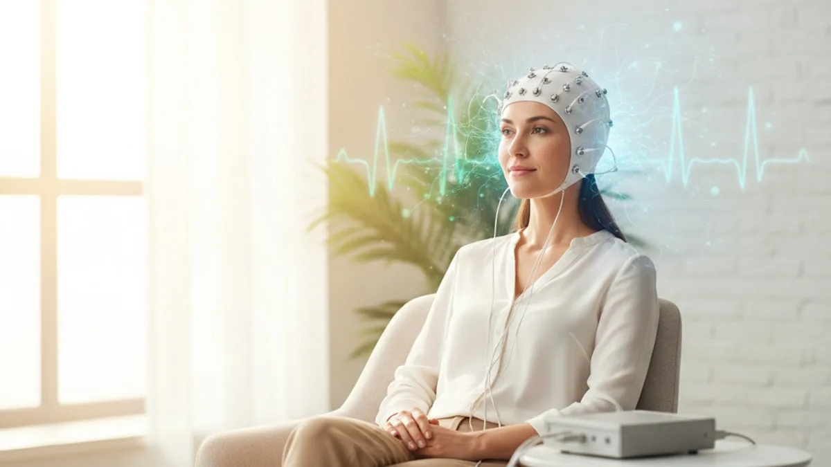
EEG Test: How Brain Wave Recording Works
A seizure lasting more than 5 minutes, a first seizure, or a person who does not wake up normally after one needs emergency care. Call 911.
What does an EEG actually record?
An EEG records tiny voltage changes generated by the brain itself, picked up by sensors resting on the surface of the scalp. Nothing is injected, nothing is swallowed, and no current is sent into the head. The machine is a listener, not a transmitter.
Those voltage changes do not come from single nerve cells firing. Research suggests that the scalp signal reflects the summed postsynaptic potentials of large, similarly oriented populations of cortical neurons acting together 1. That is why the signal is so small, measured in microvolts, and why thousands of cells have to be active in rough synchrony before anything shows up at the scalp at all.
The signal also has to travel outward through cerebrospinal fluid, skull and skin, each of which blurs and weakens it. The trade-off is worth understanding. An EEG is poor at telling you exactly where in the brain something happened, but it is extremely good at telling you when, down to the millisecond. Few other tests of brain function offer that kind of timing.
Once digitised, the recording can be filtered, re-referenced and displayed in different ways by the person reading it. Studies indicate that these analysis methods are the standard toolkit used to describe EEG signals in both neuroscience and clinical neurology 2. In practice this means the raw recording is not a single picture. It is a dataset that a trained reader examines from several angles.
Why might a clinician order one?
The most common reason is an unexplained spell, meaning a sudden episode of shaking, staring, collapse, confusion or loss of awareness that nobody can fully account for. The recording gives a clinician a window into the electrical background of the brain between episodes, and occasionally during one.
Research suggests that EEG contributes to diagnosing epilepsy in adults, to classifying the seizure type, and to ongoing monitoring decisions once a diagnosis has been made 3. It is one input among several, sitting alongside the account of what happened, a witness description, a physical examination and sometimes brain imaging.
Other reasons a recording may be requested include persistent unexplained confusion, episodes that happen out of sleep, assessment of brain activity in people who are critically unwell, and questions about whether an episodic change in behaviour has a neurological explanation. In mental health settings the test is used selectively rather than routinely. Research suggests that routine EEG in psychiatric practice is most informative for specific indications, such as episodic behavioural change or a suspected underlying neurological cause, rather than as a general screening test 4.
What an EEG does not do is measure intelligence, personality, stress levels or thinking. It cannot read thoughts, and no scalp recording can identify a psychiatric condition on its own.
What happens during the appointment?
A routine appointment has three parts, setup, recording and removal, and the person running it is usually a trained neurodiagnostic technologist rather than a doctor.
Setup starts with measuring your head and marking electrode positions, because placement follows an international system based on landmarks like the bridge of the nose and the bony bump at the back of the skull. Each site may be cleaned with a mildly gritty gel so the sensor makes good contact. Electrodes are then fixed with a water-soluble paste, or applied as a cap, and connected to the amplifier. Minimum recording standards published by international neurophysiology and epilepsy bodies describe how electrodes should be placed and how long a technically satisfactory recording needs to run 5.
Recording itself is undramatic. You lie or sit still with your eyes closed, opening and closing them when asked, breathing normally most of the time. The technologist watches the live trace, notes anything you do (a cough, a swallow, a jaw clench) and may ask you to perform the activation procedures described further down. You may be encouraged to drift into light sleep.
Removal takes a few minutes. The sensors lift off, the paste comes out with warm water, and there are no restrictions afterwards unless you were asked to stay awake the night before.
Modern systems store everything digitally. Studies indicate that standardised digital recording specifications exist precisely so the reader can re-display the same recording with different filters and montages without losing the original signal 6.
Does an EEG hurt?
No. The test is painless, and that is one of the few things about it that can be said flatly.
The sensations people actually report are mild and mechanical. The skin preparation gel feels like a firm scrub on the scalp. The paste is cold and sticky. Lying still for a while can be boring, and some people get a stiff neck. A few develop a patch of redness or a little tenderness where an electrode sat, which usually settles within a day.
Two parts of the recording can feel odd rather than painful. Deep fast breathing can make you lightheaded, tingly in the fingers or slightly short of breath, which passes within a minute of stopping. The flashing light can be visually unpleasant for people who find bright strobing uncomfortable.
There is no radiation, no needle, no scan preparations to take beforehand, and no electricity delivered to the brain. If any part of the session becomes uncomfortable, tell the technologist, because activation steps can be stopped at any point.
How should you prepare for the recording?
Start with your hair, because contact quality decides how readable the recording is. Wash it the night before or that morning with plain shampoo, then skip conditioner, oils, serums, gels, mousse, hairspray and dry shampoo. Arrive with hair dry and loose. Braids, extensions, weaves and thick product residue all get in the way of electrode contact, so tell the clinic in advance if removing them is not realistic, and they will advise.
Sleep is the other variable. For a standard recording you are usually asked to sleep normally. For a sleep or sleep-deprived recording you will be given specific instructions about how much of the night to stay awake, and you should arrange for someone else to drive you.
Eat normally unless the clinic tells you otherwise, because low blood sugar makes the session harder and can alter the background rhythm. Many clinics ask you to skip caffeine on the morning of the test, since it makes drowsiness and light sleep harder to reach.
Medicines deserve a direct word. Some medicines change the background rhythm or the chance of capturing an abnormality, so ask your prescriber well before the appointment and never stop or alter anything on your own. Bring a current list with you, plus a short written description of your episodes and, if possible, an account from someone who has witnessed one. Finally, bring a comb or a hat, because paste residue is normal and washes out at home.
What is a sleep-deprived recording and why would one be requested?
A sleep-deprived recording is a scalp EEG performed after a planned, partial night of lost sleep, so that drowsiness and light sleep occur during the session. Some electrical abnormalities surface mainly in those states and stay hidden during ordinary alert wakefulness.
Research suggests that sleep-deprived EEG has a higher diagnostic yield for epileptiform abnormalities than standard routine recording alone 7. That is the whole reason a clinician would ask you to go through an uncomfortable night. Minimum recording standards also set out how sleep recordings should be carried out so that the result is interpretable rather than just noisy 5.
Practical points matter here. Follow the clinic's instruction exactly, because staying up longer than asked is not better. Do not drive yourself to or from the appointment. Expect to feel flat for the rest of the day, and plan nothing demanding. In people with a known seizure disorder, sleep loss can itself make an event more likely, which is a reason to have the test supervised in a clinic and to have someone with you.
What are activation procedures?
Activation procedures are deliberate steps during the recording that make hidden abnormalities more likely to appear. The two most familiar are deep fast breathing for a few minutes and intermittent flashing light delivered by a lamp placed in front of the closed or open eyes. Drowsiness and light sleep count as a third.
The logic is simple. A brain that looks unremarkable at rest may reveal a pattern under a mild physiological challenge, and capturing that pattern changes what the reader can say about the recording.
There is a small chance that an activation procedure provokes a seizure in a susceptible person, which sounds alarming but is actually part of the design. It happens in a monitored room, with trained staff watching the live trace, and the procedure is stopped immediately. Deep breathing is often shortened or skipped in people with certain heart or lung conditions, and the technologist will ask about that beforehand.
If you have had an unpleasant reaction to flashing lights in the past, say so before the lamp is positioned.
Which type of recording might be used?
The type depends on how often your episodes happen and what question the clinician is trying to answer. A short recording suits a general look at the background. Capturing an actual event usually needs a longer one.
| Recording type | What it involves | Why it may be chosen |
|---|---|---|
| Routine scalp EEG | Short clinic session with sensors on the scalp, including activation procedures | First look at the electrical background after an unexplained spell |
| Sleep or sleep-deprived EEG | Same setup, performed after planned partial sleep loss so drowsiness and light sleep occur | When a routine recording was unrevealing and abnormalities may be state-dependent |
| Ambulatory EEG | Sensors stay on while you go home with a portable recorder for one or more days | Episodes are infrequent and unlikely to occur in a short clinic session |
| Video EEG monitoring | Inpatient recording with simultaneous video over an extended admission | The event itself needs to be captured and matched to the trace, second by second |
| Continuous EEG in critical care | Ongoing bedside recording in an intensive care setting | Assessing brain activity in a person who is unresponsive or critically unwell |
Longer recordings are not automatically better. They cost more, involve more disruption, and produce hours of data that still have to be reviewed by a person. The right choice is the shortest recording likely to answer the question, which is a judgement for the clinician who knows your history.
What do the brain wave bands mean?
EEG activity is conventionally divided into frequency bands, and studies indicate that these band definitions are the standard descriptive framework used when EEG signals are analysed 2. The bands describe speed, not emotion or thought.
| Band | Approximate frequency | What it commonly reflects |
|---|---|---|
| Delta | Below 4 Hz | Deep sleep in adults; prominent slow activity during wakefulness is noted by the reader |
| Theta | 4 to 8 Hz | Drowsiness and light sleep |
| Alpha | 8 to 13 Hz | Relaxed wakefulness with eyes closed, strongest over the back of the head |
| Beta | 13 to 30 Hz | Alert, active wakefulness; some medicines increase it, so ask your prescriber about anything you take |
| Gamma | Above 30 Hz | Faster activity, harder to record cleanly from the scalp because muscle signals overlap it |
A report rarely leads with band labels. It describes whether the background rhythm is present and symmetrical, whether it reacts normally when you open your eyes, whether there is slowing where there should not be, and whether any sharp or spike-like discharges appeared. Those descriptive terms carry the meaning. A single mention of theta activity in a drowsy adult says almost nothing on its own.
Can the test tell a seizure apart from a faint?
It contributes, but it does not settle the question by itself. A faint and a seizure can look similar to bystanders, because a brief faint often includes stiffening or a few jerks, and both can end with a person on the floor looking confused.
What separates them is usually the story. Clinicians ask about what came immediately before (standing for a long time, pain, heat, the sight of blood, a feeling of warmth, nausea, tunnel vision or muffled hearing), how long the person was unresponsive, whether the side of the tongue was bitten, how quickly full awareness returned, and whether there was a long period of confusion afterwards. A recording between episodes may show a pattern that supports one explanation, but a clean recording does not rule a seizure out.
Because fainting often has a circulatory explanation rather than an electrical one, the workup commonly includes a heart rhythm check and blood pressure measurements lying and standing. If this is the question in front of you, the overview of fainting causes sets out the main categories, and dizziness causes is the companion piece for people whose main symptom is lightheadedness or spinning rather than a full loss of consciousness.
Bring a witness account to the appointment if you possibly can. It is often worth more than any single test result.
What can a normal result not rule out?
A normal recording does not mean nothing is wrong, and that single sentence prevents a lot of unnecessary reassurance. An EEG captures a short window of electrical activity, and most people are not having an episode during it.
Research suggests that a first routine recording is normal in a substantial share of adults who do have epilepsy, which is why repeat recordings, sleep recordings or longer monitoring are often requested after an unrevealing first test 3. Research suggests that performing the recording after sleep deprivation raises the chance of detecting epileptiform activity compared with a standard routine recording 7.
The reverse also holds. An abnormal recording is not a diagnosis. Some patterns appear in people who never have a seizure, muscle tension and eye movement can mimic abnormalities, and the reader's job includes separating artefact from genuine brain activity.
An EEG also says nothing about brain structure. It will not show a structural lesion, a bleed or a narrowed vessel, which is what imaging is for. And it does not identify anxiety, depression or attention problems. Research suggests that routine EEG in psychiatry earns its place for defined indications rather than as a broad diagnostic screen 4.
What are the side effects of an EEG test?
Side effects are minor and short-lived, which is one reason the test is used so widely. There is no radiation and no injection.
The common ones are cosmetic or mechanical. Expect paste residue in the hair, occasional redness or mild tenderness at electrode sites, and sometimes a small patch of skin irritation from adhesive during longer monitoring. People with sensitive skin should mention it at the start so a gentler fixing method can be used. Scalp soreness is more likely after multi-day recordings than after a short session.
Deep fast breathing can cause lightheadedness, tingling in the hands or lips, and a brief feeling of breathlessness, all of which resolve quickly. Flashing light can cause visual discomfort. In people with photosensitivity, light stimulation can provoke an event, which is why it is performed under supervision and halted at the first sign of a response.
Sleep-deprived recordings carry their own trade-off. Tiredness, poor concentration and low mood for the rest of the day are expected, and in people with a seizure disorder sleep loss can raise the chance of an event, which is why transport should be arranged and company is sensible.
If an electrode site stays sore, swollen or weepy over the following days, contact the clinic.
Which blood markers are often checked around the same time?
Blood tests do not replace a recording, but basic chemistry is commonly checked after an unexplained spell, because shifts in the body's internal balance can change how the brain behaves. Sodium is one of those markers. Compare your result with the range printed on your lab report; FitScript's optimal range is 138 to 142 mEq/L, an optimization target rather than a diagnostic cut-off. Ranges vary by lab, age and sex, and the dedicated marker guide covers measurement and what a high or low result may reflect. FitScript's membership includes 150+ biomarkers tested per year, with results interpreted by Atlas, FitScript's AI health advisor. Any decision that follows belongs to a licensed provider who has reviewed the full picture.
Who reads the recording, and what does the report say?
A neurologist or clinical neurophysiologist reads the recording, not the machine and not the technologist who performed it. Automated detection software may flag segments for attention, but a trained human reviews the trace and writes the interpretation.
The report usually follows a predictable shape. It describes the background rhythm and whether it is present, symmetrical and reactive to eye opening. It notes any focal or generalised slowing. It records whether sharp waves, spikes or other epileptiform discharges appeared, and where. It states what happened during deep breathing, light stimulation and sleep. Then it gives an impression, which is the reader's summary of what the recording shows, written in careful language rather than as a verdict.
Digital storage matters here more than most people expect. Studies indicate that recording standards for digital media exist so the whole recording is preserved and can be re-reviewed with different display settings, which supports a second opinion later 6.
Turnaround varies by clinic and by recording type, so ask at the appointment how and when you will get the result, and who will explain it. The technologist will not give you an interpretation on the day, and that is normal practice rather than evasion.
When is it worth talking to a clinician?
Some situations need emergency care rather than an appointment. A seizure lasting more than 5 minutes, a first seizure, repeated seizures without full recovery in between, or a person who does not wake up normally afterwards all need emergency services. Call 911. The same applies to a collapse with chest pain, new one-sided weakness or difficulty speaking, or a head injury during the event.
Book a clinical review promptly, rather than waiting, if you have had any episode of loss of consciousness that has not yet been assessed, if episodes are becoming more frequent or longer, if you faint during exertion or while lying down, if you wake with a bitten tongue or unexplained injuries, or if confusion after an episode lasts longer than it used to. Ask about driving rules too, because restrictions after a loss of consciousness are set where you live and your clinician can tell you what applies.
Bring three things to that appointment. A written timeline of the episodes, an account from someone who has seen one, and a current list of everything you take, with any medicine questions directed to your prescriber. This article is educational and is not medical advice. Discuss your results and any treatment with a clinician or licensed provider who can review your full history.
Key takeaways
- An EEG records the brain's own electrical activity through scalp sensors. It is painless, sends nothing into the head, and uses no radiation.
- The scalp signal comes from large populations of cortical neurons acting together, which is why timing is its strength and precise location is not.
- Preparation decides data quality. Clean dry hair with no product, normal eating, and clear instructions about sleep and about any medicines from your prescriber.
- Activation procedures, deep breathing, flashing light and drowsiness, are standard parts of a routine recording, not extras.
- A normal recording does not rule out epilepsy, and an abnormal one is not a diagnosis. Repeat, sleep-deprived or longer recordings are often the next step.
- A seizure over 5 minutes, a first seizure, or failure to wake normally afterwards needs emergency care.
References
- The neurophysiological bases of EEG and EEG measurement: a review for the rest of us. Psychophysiology, 2014.
- The applied principles of EEG analysis methods in neuroscience and clinical neurology. Military Medical Research, 2023.
- Clinical utility of EEG in diagnosing and monitoring epilepsy in adults. Clinical Neurophysiology, 2018.
- A review of indications for routine EEG in clinical psychiatry. Hospital & Community Psychiatry, 1992.
- Routine and sleep EEG: Minimum recording standards of the International Federation of Clinical Neurophysiology and the International League Against Epilepsy. Epilepsia, 2023.
- American Clinical Neurophysiology Society Guideline 4: Recording Clinical EEG on Digital Media. The Neurodiagnostic Journal, 2016.
- The diagnostic value of sleep-deprived EEG in epilepsy: A meta-analysis. Seizure, 2024.
Common questions
Is an EEG test painful?
No. Sensors rest on the surface of the scalp and record activity without sending anything into the head. The main sensations are a firm scrub during skin preparation, cold sticky paste, and stiffness from lying still. Deep breathing during the session can feel lightheaded or tingly, and that passes within a minute of stopping.
How long does an EEG appointment take?
It depends entirely on the type of recording. A routine scalp recording is a single clinic session covering setup, recording and removal, while ambulatory and inpatient video recordings run for a day or longer. Ask the clinic booking you, because they will tell you the length and whether you need someone to drive you home.
Should I wash my hair before an EEG?
Yes. Wash with plain shampoo the night before or that morning, then skip conditioner, oils, serums, gel, mousse, hairspray and dry shampoo, and arrive with hair dry and loose. Product residue sits between the sensor and the scalp and degrades the recording. Tell the clinic in advance about braids, extensions or weaves so they can advise.
Does a normal EEG mean I do not have epilepsy?
No. The recording captures a short window of activity, and most people are not having an episode during it, so a normal result does not exclude a seizure disorder. That is why a clinician may request a repeat recording, a sleep-deprived recording or longer monitoring. Your description of the episodes and a witness account carry a lot of weight alongside the test.
Can an EEG detect anxiety, depression or ADHD?
No. The test describes the electrical rhythms of the brain, not mood, attention, personality or thinking. In mental health care it is used selectively, usually when there is a question about an underlying neurological cause for an episodic change in behaviour. A clinician decides whether it is appropriate in a given situation.
Should I stop my medicines before the test?
Do not change anything on your own. Some medicines can alter the recording, so ask your prescriber well before the appointment and follow their instruction exactly. Bring a current list of everything you take to the appointment so the person reading the recording knows what was on board.
Can I drive after an EEG?
After a standard recording there are usually no restrictions from the test itself. After a sleep-deprived recording you should arrange for someone else to drive you both ways, because you will be tired and your concentration will be poor. Driving rules following a loss of consciousness or a seizure are set where you live, so ask your clinician what applies to you.
What is the difference between an EEG and a brain scan?
An EEG measures function over time, showing the brain's electrical rhythms moment by moment. A brain scan shows structure, such as the shape of tissue or a lesion. They answer different questions, and a clinician may request one, the other or both depending on what needs clarifying.