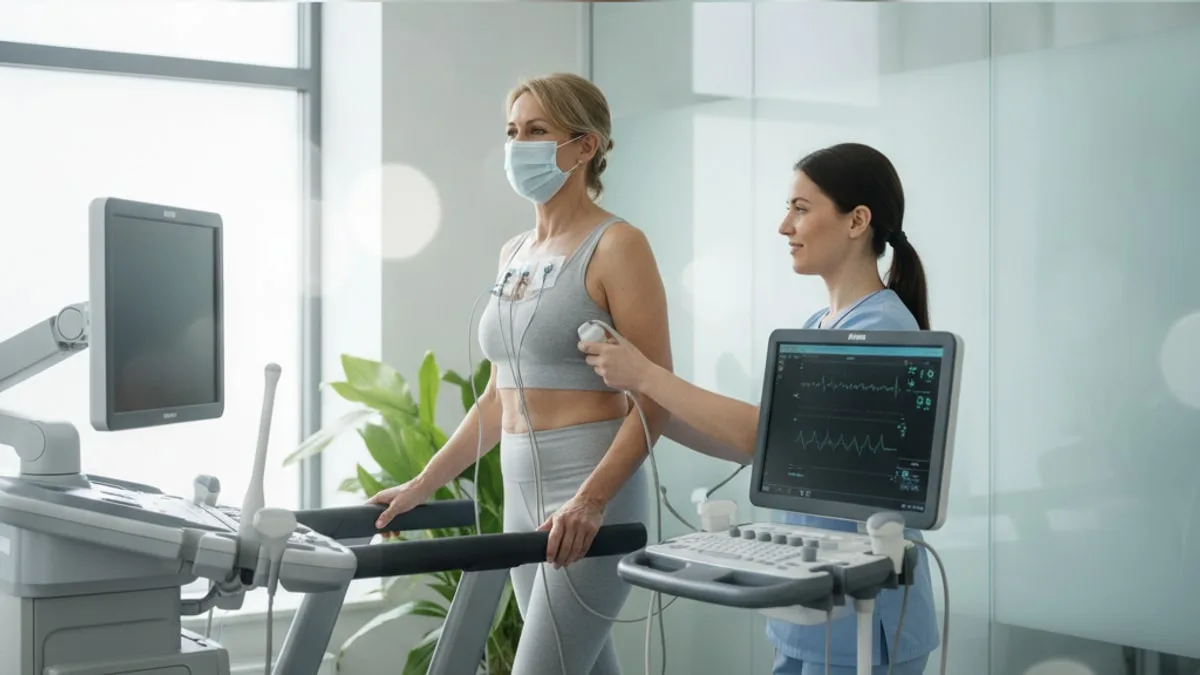
Stress Echocardiogram: Heart Ultrasound During Exercise
Chest pain at rest, chest pain that will not settle, pain spreading to the arm, jaw or back, sudden severe breathlessness, fainting, or collapse with a racing heartbeat are emergency signs. Call 911 or go to the emergency department.
What is this test in plain terms?
It is an ultrasound scan of the heart performed twice in one appointment, once with the heart at rest and once with the heart working hard, so the two sets of pictures can be placed side by side and compared.
A sonographer or clinician holds a probe against the chest wall and sound waves build moving pictures of the beating heart. Those pictures show the four chambers, the thickness and movement of the muscle walls, the valves opening and closing, and the direction and speed of blood flowing through them. At rest, a set of standard views is recorded and stored digitally so the same views can be captured again under stress and displayed together, usually as a split screen of matched loops.
The stress itself can come from the body or from a medicine. Walking on a treadmill, pedalling an upright or a semi-reclined bicycle, or a medicine given under supervision all raise the work the heart has to do. Throughout the appointment the heart rhythm is traced on an ECG and blood pressure is checked at intervals, so the images sit alongside a record of heart rate, rhythm, blood pressure and any symptoms.
The comparison is the point. A single still frame of the heart is far less informative than the same segment of muscle photographed calm and then photographed under demand. Research suggests the rest and stress comparison is what makes the scan useful for detecting reduced blood supply in coronary disease 1, and exercise echocardiography has been used in this way since digital image capture made side by side review practical 2.
Why does pushing the heart reveal what a resting scan can miss?
Because a narrowed coronary artery can still deliver enough blood when the heart is idling, and only falls short when demand climbs.
The heart muscle is fed by its own arteries. When plaque narrows one of them, flow through the narrowing can remain adequate for a resting heart beating slowly against a modest blood pressure. Raise the heart rate, raise the force of each contraction and raise blood pressure, and the muscle downstream of the narrowing needs more oxygen than the artery can deliver. That mismatch does not announce itself all at once. The affected segment of muscle first relaxes less well, then contracts and thickens less than its neighbours, and those movement changes can be seen on ultrasound. Electrical changes on the ECG and chest discomfort, if they appear at all, tend to come later in that sequence.
This is why imaging adds information to an exercise ECG. The ECG records the electrical consequence of reduced supply across the whole heart; the images show which part of the muscle is struggling and at what workload. A new or worsening movement abnormality in a defined region during stress is the finding the scan is built around 1.
The same logic extends past the coronary arteries. Valves behave differently under load, pressures in the lung circulation rise with exercise, and the pumping reserve of a weakened heart only reveals itself when the heart is asked for more. A 2024 clinical consensus statement from the European Association of Cardiovascular Imaging describes use of the technique in chronic coronary syndromes and beyond coronary artery disease, including a multiparametric approach that looks at wall motion alongside measures such as contractile reserve, lung ultrasound signs, coronary flow velocity reserve and heart rate reserve 3.
What happens from arrival to the final images?
The appointment runs as one continuous sequence of resting images, stress, peak or immediate post-stress images, and recovery monitoring.
After check in, the clinical team confirms your history, your symptoms and the reason the scan was requested, and explains what they plan to do. Small electrodes are placed on the chest for the ECG, and a blood pressure cuff goes on an arm. You then lie on your left side while the resting images are recorded in a set of standard views. Those views matter more than they look; the whole comparison depends on capturing the same windows again under stress.
If exercise is the stressor, you move to a treadmill or a bicycle. Workload rises in stages until you reach a target heart rate, until symptoms or a change in rhythm or blood pressure prompt the team to stop, or until you are too tired to continue. On a semi-reclined bicycle, images can be captured while you are still pedalling, so the heart is photographed at peak load. On a treadmill, you return to the couch the moment you stop and images are recorded quickly, because movement abnormalities can fade within a short window after exercise ends 2.
If a medicine supplies the stress, you stay on the couch. Heart work is raised in supervised steps while images are recorded at each stage, which allows continuous imaging through the whole range of workloads 4.
Monitoring continues into recovery until heart rate, blood pressure and rhythm settle back toward where they started. The images are then reviewed, scored and reported, and the report goes to the clinician who asked for the scan.
How is the heart stressed when walking on a treadmill is not an option?
By a medicine given under supervision, or, in selected people who already have a pacemaker, by raising the heart rate through the device.
Plenty of adults cannot exercise hard enough for a useful scan. Joint pain, previous surgery, lung disease, balance problems, deconditioning and neurological conditions all cap how much work the legs can do before the heart reaches the workload the test needs. Medicine-driven stress solves that problem in two broad ways. One approach raises the force and rate of contraction so the muscle demands more oxygen, much as exercise does. The other alters the distribution of flow in the coronary circulation so that regions fed by a narrowed artery receive relatively less, which makes the difference visible on imaging 4. Which approach is used, and whether either is suitable for you, is a decision for the supervising clinician.
Pacing is a third route. In people who already have a pacemaker, the heart rate can be raised stepwise through the device without any exercise and without a medicine, and pacing stress echocardiography has been described as an option in this group 5.
One practical note worth raising when you book. Some medicines you already take can blunt the heart rate response and make a stress scan harder to interpret, and some are paused before certain tests. Do not change anything on your own. Ask your prescriber what to do on the day, and bring your current list with you.
What is wall motion, and what are the readers of the scan looking for?
Wall motion is how each segment of heart muscle thickens and moves inward with every beat, and the reader is looking for any segment that moves less well under stress than it did at rest.
The left ventricle is divided into named segments, each roughly mapped to the territory of one of the main coronary arteries. At rest, each segment is graded on how it moves. The same grading is repeated on the stress images. Four broad patterns come out of that comparison. A segment that moved normally and still moves normally under stress. A segment that moved normally at rest and moves poorly under stress, which is the classic sign of flow that cannot keep up with demand. A segment that moved poorly at rest and improves with low level stress, which can reflect muscle that is still viable. And a segment that moved poorly at rest and does not change, which may reflect older damage 1.
Readers do not stop at wall motion. Overall pumping function, chamber size, valve opening and leakage, flow velocities and estimated pressures in the lung circulation are all compared between the two states, and the consensus approach adds reserve measures to that list rather than relying on wall motion alone 3. How much workload you achieved, your symptoms, your blood pressure response and your ECG are read alongside the pictures, because a scan that stopped at a low heart rate answers a narrower question than one that reached target.
Related: Nuclear Stress Test: What It Shows and What to Expect
What can a normal result and an abnormal result each suggest?
A normal result at an adequate workload points away from flow-limiting coronary narrowing; an abnormal result points toward a region of muscle that is not getting what it needs under load, and neither outcome is a diagnosis on its own.
Take them in turn. When every segment thickens and moves normally at a workload that genuinely stressed the heart, the scan has not found the pattern that reduced coronary flow produces. A normal result in this setting is associated with a lower likelihood of significant flow-limiting disease, and the 2024 consensus statement sets out how results of this kind are used for risk assessment as well as diagnosis 3. That is useful information, and it is not a promise about the future. It does not say that plaque is absent, and it does not replace attention to blood pressure, blood markers, body composition, sleep, activity and smoking status.
When a segment or a group of segments moves less well under stress, the observation is that part of the muscle underperformed when asked for more. What that may reflect is reduced flow in the artery supplying that territory, and the report usually notes which territory, how many segments, and at what workload the change appeared, because extent and threshold both shape what comes next. The next step is a conversation with the clinician who ordered the scan, who weighs the images against your symptoms, your other test results and your history. That conversation might lead to further imaging, to an invasive look at the coronary arteries, or to changes managed entirely without a procedure. Treatment decisions belong to a licensed provider.
Results sit between the two extremes more often than guides admit. Images can be equivocal, a scan can stop below target heart rate, and a borderline finding can be reported as inconclusive. An inconclusive scan is not a wasted scan; it tells the clinician that a different test may answer the question better.
What does the scan add when a valve is the question?
It shows how a valve behaves when flow increases, which is often where resting measurements and real-life symptoms stop agreeing.
Valve disease is graded at rest, but people live under load. Someone with a tight or leaking valve may have resting numbers that look moderate and symptoms that look severe, or resting numbers that look severe while they insist they feel fine. Studies indicate that stress testing with ultrasound helps clarify these mismatches in valvular heart disease by showing gradients, leakage and pressures as the heart works harder 6.
Three situations come up repeatedly. First, a narrowed aortic valve in a heart that is not pumping strongly can produce low gradients that understate how tight the valve really is; low level medicine-driven stress is used to distinguish truly severe narrowing from apparent severe narrowing caused by weak flow 7. Second, leakage across a valve can worsen with exertion in a way a resting scan never captures, along with a rise in pressure in the lung circulation 7. Third, in people who report no symptoms despite severe valve disease, exercise with imaging can show a limited exercise capacity or an abnormal blood pressure response that reframes the whole picture 6.
None of this decides surgery or any other procedure. It gives the heart team one more piece of information than a resting scan can provide, and the decision stays with the clinicians caring for the person.
What does it add when breathlessness is the main complaint?
It can show whether the heart is the reason exertion feels hard, by measuring pumping reserve, filling pressures, valve behaviour and lung circulation pressures during or immediately after effort.
Breathlessness on exertion has many possible explanations, and a resting scan that looks unremarkable does not close the question. Research suggests that, in heart failure, stress echocardiography contributes information on contractile reserve, filling pressures and dynamic valve leakage that resting imaging does not provide 8. Contractile reserve means how much more the heart can squeeze when pushed. Filling pressure assessment during exertion is sometimes described as diastolic stress testing, and it addresses a specific question about whether pressures inside the heart rise abnormally when the heart is asked to fill faster.
The technique is also applied outside coronary disease to heart muscle conditions and to the pulmonary circulation, and joint recommendations from the European Association of Cardiovascular Imaging and the American Society of Echocardiography describe its use in non-ischaemic heart disease 9.
For an adult who simply knows that stairs have become harder over the last year, the practical value is sorting. The result may point toward the heart, toward the lungs, toward conditioning, or toward something else entirely, and it gives the clinician a reason to look in one direction rather than guessing. What it cannot do is replace the history. How quickly the change came on, what makes it worse, whether it happens lying flat and whether swelling came with it all stay central.
When is ultrasound chosen instead of a nuclear scan?
The choice depends on the clinical question, how clear the ultrasound pictures are likely to be, whether the person can exercise, and what the local service offers.
Both approaches stress the heart and image the result; they image different things. Ultrasound shows muscle movement, chamber size, valves and pressures. Nuclear imaging shows how blood is distributed through the muscle at rest and under stress. Research suggests that detection of coronary disease with ultrasound during stress compares favourably with other established imaging approaches 1.
| Feature | Stress echocardiography | Nuclear stress imaging | Exercise ECG without imaging |
|---|---|---|---|
| What it shows | Muscle movement, chamber size, valves, estimated pressures | Distribution of blood flow in the heart muscle | Electrical changes, symptoms, exercise capacity |
| Imaging method | Ultrasound, no ionising radiation | Radiation-based imaging | No imaging |
| Stress used | Exercise, medicine-driven stress, or pacing in selected people | Exercise or medicine-driven stress | Exercise only |
| Extra information it can give | Valve behaviour under load, pumping reserve, lung circulation pressures | Flow comparison between rest and stress across territories | Heart rate and blood pressure response, workload achieved |
| Main practical limit | Picture quality depends on the ultrasound window and on operator skill | Radiation exposure and a longer appointment | Cannot show which region of muscle is affected |
Some people have ultrasound windows that are hard to image, because of chest shape, body composition or lung disease, and in that case a test that does not depend on acoustic access may answer the question better. Where imaging is feasible and the question involves valves, pumping reserve or exertional breathlessness, the ultrasound approach brings information no perfusion map contains. The decision is not yours to make alone and it is not a ranking of better and worse tests; it is a match between question and tool, made by the requesting clinician.
How should you get ready for the appointment?
Follow the written instructions your clinic sends, because preparation differs by the type of stress used, and bring a current list of everything you take.
A few things are close to universal. Wear clothes and shoes you can walk or pedal in, since exercise appointments are real exercise. Expect electrodes on bare skin on the chest. Allow more time than the scan itself takes, because monitoring continues after stress ends. Bring reports from any previous heart imaging if they were done elsewhere, as a prior comparison makes a new one easier to read.
The variable parts are food, drinks and medicines. Some clinics ask for a period without food before the appointment; some ask you to avoid caffeine-containing drinks for a set time before a medicine-driven test. Those instructions come from the department performing the scan, so follow the ones you were given rather than general advice. On medicines, some medicines you take can change the heart rate response; ask your prescriber whether to take them on the day and never stop anything by yourself.
One more preparation worth knowing about. When the borders of the heart muscle are hard to see, the team may use a scan preparation to sharpen the outline so each segment can be judged properly. If they plan anything of that kind, they will explain it and ask for your consent first. Ask whatever you want to ask before the scan starts; the team would rather answer in advance than mid-exercise.
What are the risks, and how is the appointment kept safe?
Stress deliberately provokes the heart, so the appointment is supervised throughout with continuous rhythm monitoring and staff who can stop the test at any point.
During exercise, expect breathlessness, a fast heartbeat and tired legs, because that is the test working. Chest discomfort, dizziness, an abnormal blood pressure response or a rhythm change can also occur, and any of them is a reason for the team to stop early. Medicine-driven stress can bring short-lived flushing, headache, nausea, palpitations or chest discomfort while the medicine is active. Serious complications can occur with any form of cardiac stress testing, which is exactly why these appointments happen in a monitored setting with resuscitation equipment and trained staff present. The supervising clinician reviews your history first and explains the specific risks in your situation before you consent.
There is no ionising radiation involved in the ultrasound itself, which is one reason it suits people who need repeated assessment over time.
Say something the moment you feel it. Pain, lightheadedness, nausea or a feeling that something is wrong is information the team needs in real time, not after the scan. Stopping a test early is a normal event, not a failure, and the images captured up to that point are often still useful. After the appointment, if chest pain comes on at rest, does not settle, or arrives with breathlessness, sweating or fainting, call 911 or go to the emergency department.
What are the limits of what these images can answer?
The scan describes how the heart functions under load; it does not map the inside of the coronary arteries and it does not forecast an individual's future.
Several limits are worth holding onto. Picture quality varies between people and the reading depends on the experience of the person interpreting it, a limitation noted in reviews of the technique 1. A scan that stops well below target heart rate can miss a change that would have appeared at higher workload. When narrowing affects all three main arteries fairly evenly, the whole muscle can underperform together, which is harder to spot than one lagging region. And wall motion can look abnormal for reasons other than coronary flow, including previous muscle damage, conduction abnormalities and some heart muscle conditions, which is why the consensus approach reads several parameters together 3.
The flip side matters too. A normal scan is not a clean bill of health for the arteries. It means no flow-limiting pattern appeared at the workload achieved on that day. Plaque that is not yet restricting flow produces no wall motion change and still deserves attention. That is the gap blood markers and risk assessment fill, and it is the reason imaging and bloodwork are ordered together rather than as alternatives.
Where do blood markers like cholesterol fit alongside imaging?
Imaging shows how the heart performs today, while blood markers describe conditions that shape the arteries over years, so the two answer different questions and neither substitutes for the other.
LDL Cholesterol is one of the markers discussed in that conversation. FitScript's optimal range for LDL Cholesterol is 50 to 100 mg/dL, and your laboratory's own range appears as printed on your lab report. Optimal ranges are optimization targets, not diagnostic cut-offs.
What the marker measures, how it is tested, what affects it and what a result may reflect are covered in full on the LDL Cholesterol page linked above. Bring both the imaging report and the bloodwork to the same appointment, because a clinician reading them together sees more than either one alone.
Which other tests are often arranged around it?
A resting ECG, resting echocardiography, blood pressure measurement and a set of blood markers usually sit around a cardiac workup, with further imaging added when the question stays open.
On the imaging side, a resting echocardiogram often comes first and provides the baseline the stress images are compared against. Depending on the question, a clinician may add CT imaging of the coronary arteries to look at anatomy, cardiac MRI for tissue characterisation, an ambulatory rhythm recording if palpitations or fainting are part of the story, or an invasive look at the arteries. Each answers something different, and the stress scan frequently determines which one is worth doing.
On the laboratory side, a lipid panel, markers of inflammation such as CRP, a natriuretic peptide such as BNP when breathlessness is the leading symptom, blood sugar markers, kidney function and thyroid function are the kinds of labs a clinician may request around a cardiac assessment. Those are described here in words rather than numbers, because what a given value means depends on the person, the units your laboratory uses and the range printed on your own report. FitScript's membership includes 150+ biomarkers tested per year with interpretation by Atlas, its AI health advisor, and any treatment is decided by a licensed provider after reviewing the member.
The pattern across repeated panels usually carries more signal than a single draw. A marker drifting steadily in one direction over several tests tells a clinician something a one-off number cannot.
When should you talk to a clinician?
Bring any new or changing exertional symptom to a clinician, and treat chest pain at rest as an emergency rather than something to monitor.
Call 911 or go to the emergency department for chest pain at rest, chest pain that does not settle, pain spreading to the arm, jaw, neck or back, sudden severe breathlessness, fainting, or collapse with a racing heartbeat. Those signs need emergency care, not a scheduled appointment.
Arrange a prompt appointment, rather than waiting, for chest tightness or pressure that comes on with effort and eases with rest, breathlessness that has worsened over weeks or months, new swelling in the ankles or legs, palpitations with lightheadedness, or exercise capacity that has dropped without an obvious explanation. Those are the symptoms that commonly lead to a stress scan being requested in the first place.
If you already have a report in hand, read it with the clinician who ordered it. Ask what workload you reached, whether the scan was adequate for the question, which segments were described, and what the finding changes about your plan. A value or a finding outside the range described on your report is worth discussing with a clinician promptly, and nothing in a report should be ignored because it looks minor. This page is educational and not medical advice, and your results and any treatment should be discussed with a clinician or licensed provider who knows your history.
Key takeaways
- The scan images the heart at rest and under stress, then compares the two, which is where the information comes from.
- Stress can come from a treadmill, a bicycle, a medicine given under supervision, or pacing in selected people who already have a pacemaker.
- The core finding is a segment of heart muscle that moves and thickens less under load than it did at rest.
- It is used beyond coronary disease, including valve assessment and exertional breathlessness, where resting measurements and symptoms disagree.
- It uses no ionising radiation, and its main practical limits are picture quality, operator experience and the workload actually reached.
- A normal result does not rule out plaque that is not yet restricting flow, which is why blood markers stay part of the picture.
- Chest pain at rest means call 911 or go to the emergency department, not wait for a scheduled test.
References
- Stress echocardiography. Heart (British Cardiac Society), 2003.
- Exercise echocardiography. The American journal of cardiology, 1992.
- The clinical use of stress echocardiography in chronic coronary syndromes and beyond coronary artery disease: a clinical consensus statement from the European Association of Cardiovascular Imaging of the ESC. European heart journal. Cardiovascular Imaging, 2024.
- Stress echocardiography. Current pharmaceutical design, 2005.
- Pacing stress echocardiography. Cardiovascular ultrasound, 2005.
- The Role of Stress Echocardiography in Valvular Heart Disease. Current cardiology reports, 2022.
- Stress echocardiography in patients with native valvular heart disease. Heart (British Cardiac Society), 2018.
- Clinical Application of Stress Echocardiography in Management of Heart Failure. Heart failure clinics, 2020.
- The clinical use of stress echocardiography in non-ischaemic heart disease: recommendations from the European Association of Cardiovascular Imaging and the American Society of Echocardiography. European heart journal. Cardiovascular Imaging, 2016.
Common questions
What is a stress echocardiogram used for?
It is used to see how the heart muscle, chambers and valves behave when the heart is made to work harder, compared with how they behave at rest. Clinicians request it for symptoms such as chest discomfort on exertion or breathlessness that has worsened, and to help clarify valve disease when resting measurements and symptoms do not match. The result is one piece of information that the requesting clinician reads alongside your history and other tests.
Is it the same as a regular echocardiogram?
No. A regular echocardiogram images the heart at rest only. A stress echocardiogram records those resting images and then repeats the same views while or immediately after the heart is pushed to work harder, so the two sets can be compared directly.
What happens if I cannot walk on a treadmill?
The heart can be stressed without exercise. A medicine given under supervision can raise the work the heart does while you stay on the couch, and in some people who already have a pacemaker the heart rate can be raised through the device. The supervising clinician decides which approach suits your situation.
How long does the appointment take?
The length varies by clinic and by the type of stress used, so ask the department when you book. Allow extra time beyond the scan itself, because monitoring continues after the stress ends until heart rate, rhythm and blood pressure settle.
How should I prepare?
Follow the written instructions from the department performing the scan, since preparation differs by the type of stress used. Wear clothes and shoes you can exercise in, bring a current list of everything you take, and bring any previous heart imaging reports. Ask your prescriber whether to take your usual medicines on the day, and never stop anything on your own.
Does the test involve radiation?
The ultrasound itself does not use ionising radiation. That is one reason it suits people who may need repeated assessment over time. A nuclear stress test is a different type of imaging and does involve radiation exposure.
What does an abnormal result mean?
It means a part of the heart muscle moved less well under load than it did at rest, which may reflect blood flow that cannot keep up with demand in the area supplied by one artery. It is an observation, not a diagnosis, and it is read alongside your symptoms, your other results and the workload you reached. The clinician who ordered the scan explains what it changes for you.
Who decides what happens next after the scan?
The clinician who requested the test, after reviewing the report with the rest of your picture. Next steps might include another type of imaging, a closer look at the coronary arteries, or changes managed without any procedure. Any treatment decision belongs to a licensed provider who knows your history.